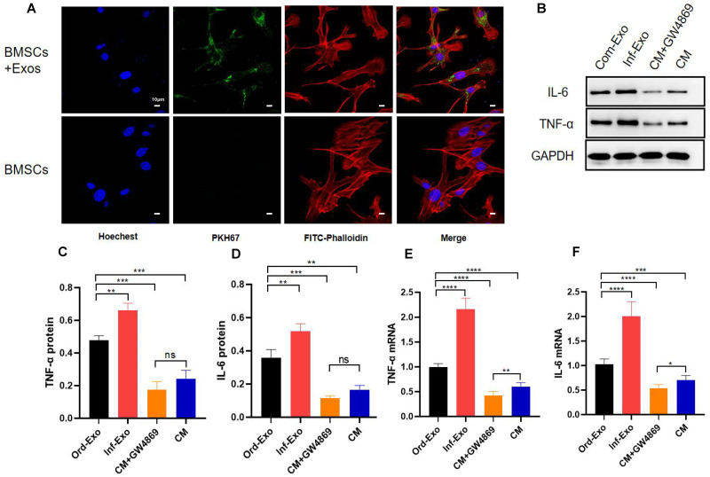
Figure 2. Exosome uptake by BMSCs and exosomes mediating the inflammatory response.
(A) Images of exosome uptake by BMSCs in fluorescence confocal microscopy. BMSCs nuclei were stained with Hoechst (blue), BMSCs Cytoskeleton were stained with FITC-Phalloidin (red), macrophage-derived exosomes were labelled with PKH67 (green). (B–D) The protein expression of TNF-α and IL-6. (E–F) The mRNA expression levels of TNF-α and IL-6. An asterisk (*) indicates P < 0.05, two asterisk (**) indicates P < 0.01, three asterisks (***) indicate P < 0.001, four asterisks (****) indicate P < 0.0001, ns indicate P > 0.05, ns indicates no significant difference.

