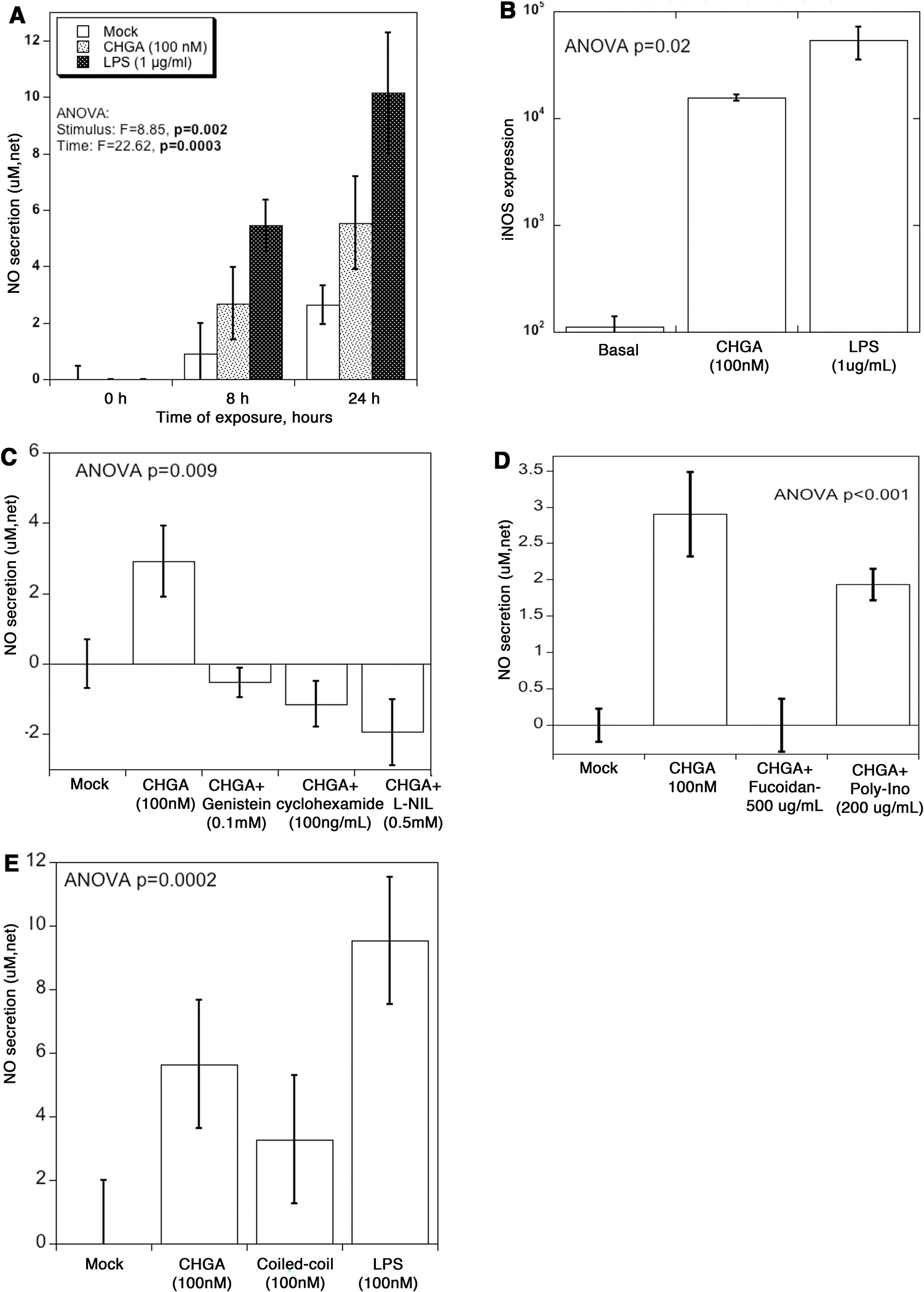
Figure 5. CHGA triggers NO release from mouse glomerular mesangial cells.

(A) CRL-1927 cells were treated with CHGA (100 nM) or LPS (1 μg/mL) for 8 and 24 hours, and the tissue culture media collected to measure total NO. The assay determined NO concentration based on the enzymatic conversion of nitrate to nitrite by nitrate reductase, followed by a colorimetric detection by Griess reagent. Control media was from cells left untreated. Net secretion was calculated by subtracting zero-hour basal secretion from all other time points. (B) Cells were treated with CHGA or LPS for 24 hours, and the total RNA extracted and processed for real-time PCR. Relative amount of the iNOS mRNA was quantified as iNOS/beta actin ratio. (C) Specificity of CHGA induced NO secretion. Cells were treated with CHGA in presence and absence of L-NIL, cycloheximide and genistein for 24 hours, and net NO secretion was measured from culture media. (D) Cells were pre-treated with SR-A blocker fucoidan or poly-ionosine for 1 hour prior to the addition of CHGA. After 24 hours tissue culture supernatant was harvested and the released NO measured. (E) Cells were treated with full length CHGA, a coiled-coil protein tropomyosin, poly-L-glutamic acid or LPS for 24-hour and the tissue culture supernatant collected to measure total NO.
