Figure 5.
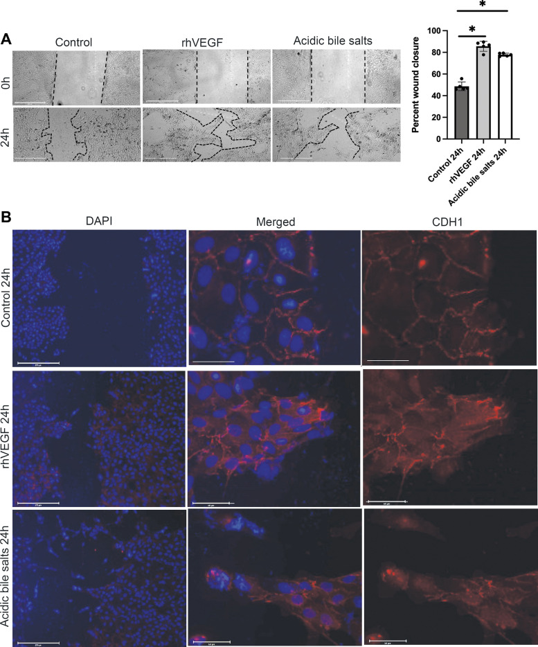
Primary cultures of Barrett’s epithelial cells treated with acidic bile salts increase collective cell migration associated with loss of membranous of CDH1. A: representative phase contrast images of scratch wounds and quantification of wound closure in BAR-B24 cells at time 0 and 24 h after treatment with recombinant human (rh)VEGF or acidic bile salts. Scale bar = 275 µm. Bar graphs depict the means ± SD from four technical replicates. *P ≤ 0.05 compared with nontreated controls; one-way ANOVA. Experiments in BAR-B24 were repeated three times. B: representative fluorescent microscopy images of wound closure 24 h after treatment with rhVEGF or acidic bile salts demonstrating the migration of cohorts of cells forming finger-like protrusions (i.e., collective cell migration). Scale bar = 275 µm. Middle and right panels are magnified images (Scale bar = 50 µm) of cell cohorts at the leading edge of the finger-like protrusions demonstrating complete or partial loss of membranous CDH1 staining following treatment with rhVEGF or acidic bile salts. CDH1, E-cadherin; rhVEGF, recombinant human vascular endothelial growth factor.

