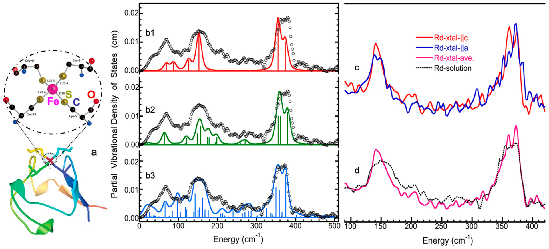
Figure 13.

(a) The crystal structure of rubredoxin (bottom, PDB: 1CAD) and the Fe site inside rubredoxin and proximal cysteine chains (top, main atoms only); (b) the observed NRVS spectra of the oxidized rubredoxin protein (open circles) vs. the NMA simulations assuming an Fe(SC)4 structure (b1); an Fe(SCC)4 structure (b2) and an Fe(SX5)4 structure (b3) [5]; (c) a single-crystal NRVS with incident X-ray parallel to a (blue) and to c (red); (d) the averaged single-crystal NRVS (purple) vs. the solution NRVS (dashed black).
