
Figure 15.
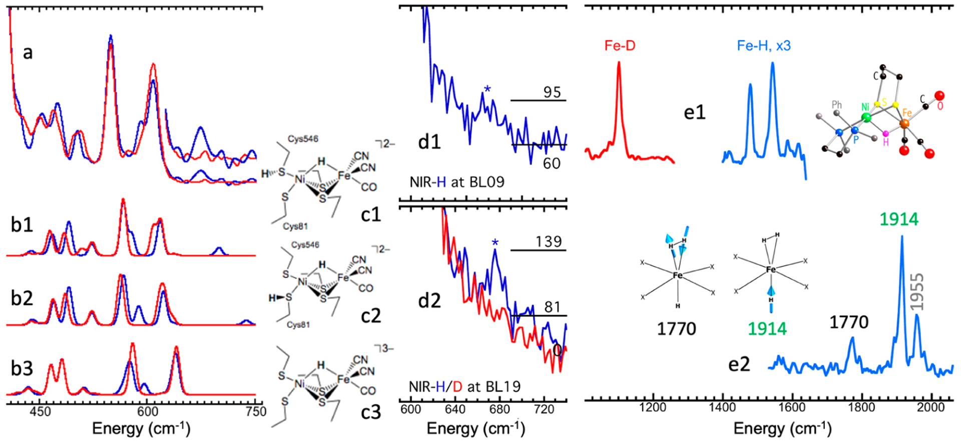
(a) NRVS for DvMF NiR–H/D (from Figure 1b); (b1–b3) DFT calculation on NiR–H/D assuming the structures in (c1–c3); (d1) the historical discovery of first Fe–H bending mode (raw NRVS) in a biological sample (DvMF hydrogenase); (d2) confirmation of the Fe–H bending (wagging) mode at higher flux BL19LXU; (e1) NRVS for the Fe–D/H stretching modes in an NiFe hydrogenase model complex (with model structure at the right); (e2) NRVS for various Fe–H stretching modes in HFe(H2) complex (with two vibrational modes at the left).