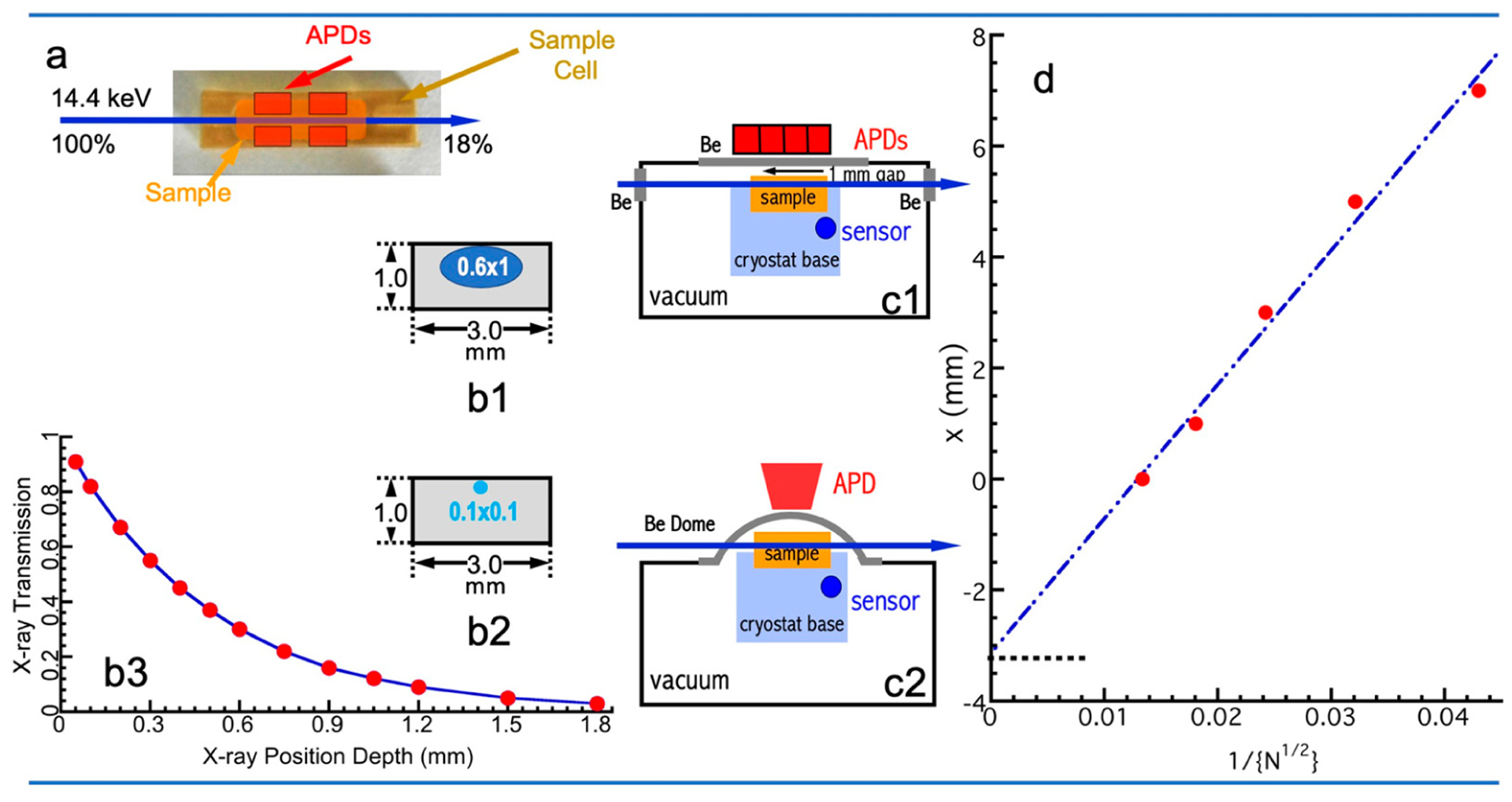
Figure 5.

(a) Geometric relationship among the >10 mm long sample/sample cell, incident X-ray beam, and APD detector array from the top view; (b) the relationship between X-ray beam and sample from the front view (cross-section) with a beam size of 1 × 0.6 mm2 (b1) and 0.1 × 0.1 mm2 (b2); (b3) the 6.4 keV X-ray transmission rate at different depth under an H2O sample; (c) side view of the incident beam, sample, and APDs: (c1) with a flat beryllium window in cryostat between the sample and the APDs; (c2) with a beryllium dome in cryostat; (d) the linear fit to the data of nominal sample-APD distance (x) vs. 1/(N1/2), where N is the nuclear resonant signal.
