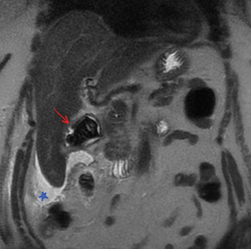
Fig. 1.

Coronal T2 single-shot fast spin echo (SSFSE)-weighted magnetic resonance imaging (MRI) of the abdomen demonstrating small amount of hyperintense ascites surrounding the liver (blue star), and a large heterogeneous, predominantly T2 hypointense signal likely within the duodenal bulb which represents a very large gallstone (red arrow; identified on imaging retrospectively only after endoscopy).
