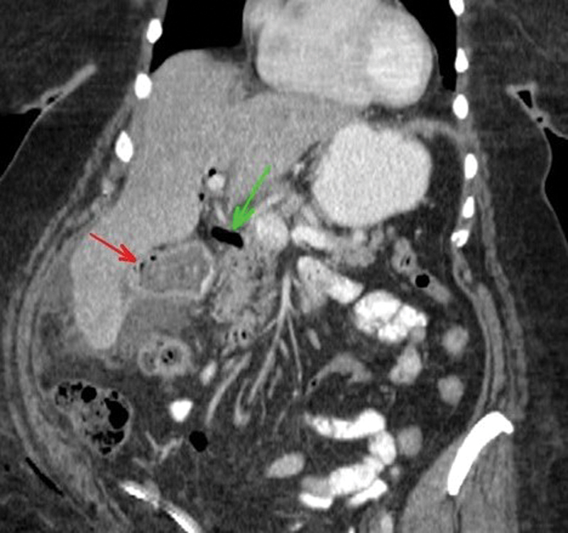
Fig. 2.

Coronal, orally- and intravenously enhanced computed tomography (CT) of the abdomen demonstrates a large calculus (red arrow) with a few foci of air as well as air within the biliary system (green arrow), suggesting cholecystoenteric fistula. A small amount of ascites surrounds the liver. Subcutaneous air within the right abdominal wall and adjacent to the gluteal muscles due to recent laparoscopy is noted.
