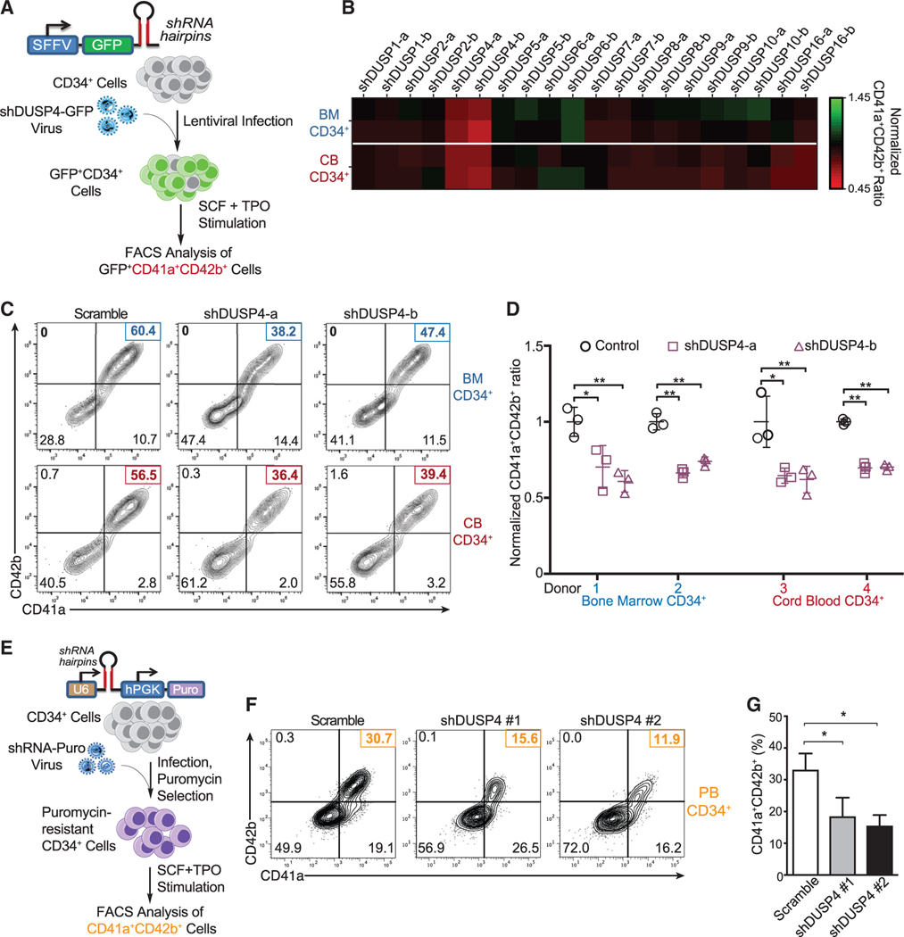
Figure 1. Identification of DUSP4 for optimal Mk differentiation.
(A) Schematic of shRNA-based screening assay to identify essential DUSPs for Mk-induced Mk differentiation. Human CD34+ cells infected with lentiviruses expressing shRNAs against DUSPs were cultured for Mk differentiation.
(B) Heatmap of the percentages of CD41a+CD42b+ cells upon DUSP knockdown on day 8. Fold changes were normalized to the percentage of double-positive cells with the group treated with control shRNA.
(C) Representative flow chart of FACS analysis of Mk differentiation using BM cells (top panel) and CB cells (bottom panel) cultured in TPO-containing medium.
(D) Summary of FACS analysis. Statistics are based on the data of three independent experiments (n = 3) with the bone marrow (BM) or cord blood (CB) cells from two donors. Data are shown as mean ± SD. Two-tailed paired t test, *p ≤ 0.05, **p ≤ 0.01.
(E) Schematic description of Mk differentiation of human peripheral blood-derived CD34+ cells with DUSP4 knockdown.
(F and G) Representative and complete FACS analysis of CD41a and CD42b markers for Mk differentiation with peripheral blood CD34+ cells upon DUSP4 knockdown on day 7. Representative plots (F) and statistics (G) are shown (n = 3, independent experiments). Data are shown as mean ± SD. Two-tailed paired t test, *p ≤ 0.05, **p ≤ 0.01.

