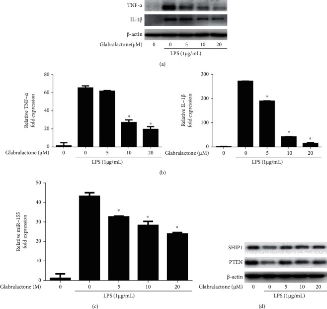
Figure 4.

Effect of glabralactone on LPS-induced proinflammatory cytokine and miR-155 expressions. The proteins (a) and mRNA (b) levels of TNF-α and IL-1β were determined by Western blotting and qRT-PCR, respectively. Cells were stimulated with LPS (1 μg/mL) in the presence of glabralactone for 4 h and was further analyzed as described in Materials and Methods. Data are representative of three separate experiments. β-Actin was used as an internal standard. (c) Effect of glabralactone on the expression of miR-155 in LPS-stimulated RAW264.7 cells. The cells were stimulated with LPS in the presence of glabralactone for 8 h. The expression of miRNA-155 was determined by qRT-PCR as described in Materials and Methods. The values are expressed as the means ± SD. (b, c) ∗p < 0.05 was considered statistically significant. (d) Effect of glabralactone on the expression of SHIP1 and PTEN in macrophage cells. Cells were treated with glabralactone for 30 min and then stimulated with LPS for 30 min. Proteins were analyzed by Western blotting. Data are representative of three separate experiments. β-Actin was used as an internal standard. Glabralactone was dissolved in 100% DMSO, and the final concentration of DMSO was adjusted to 0.1%.
