
Fig. 3.
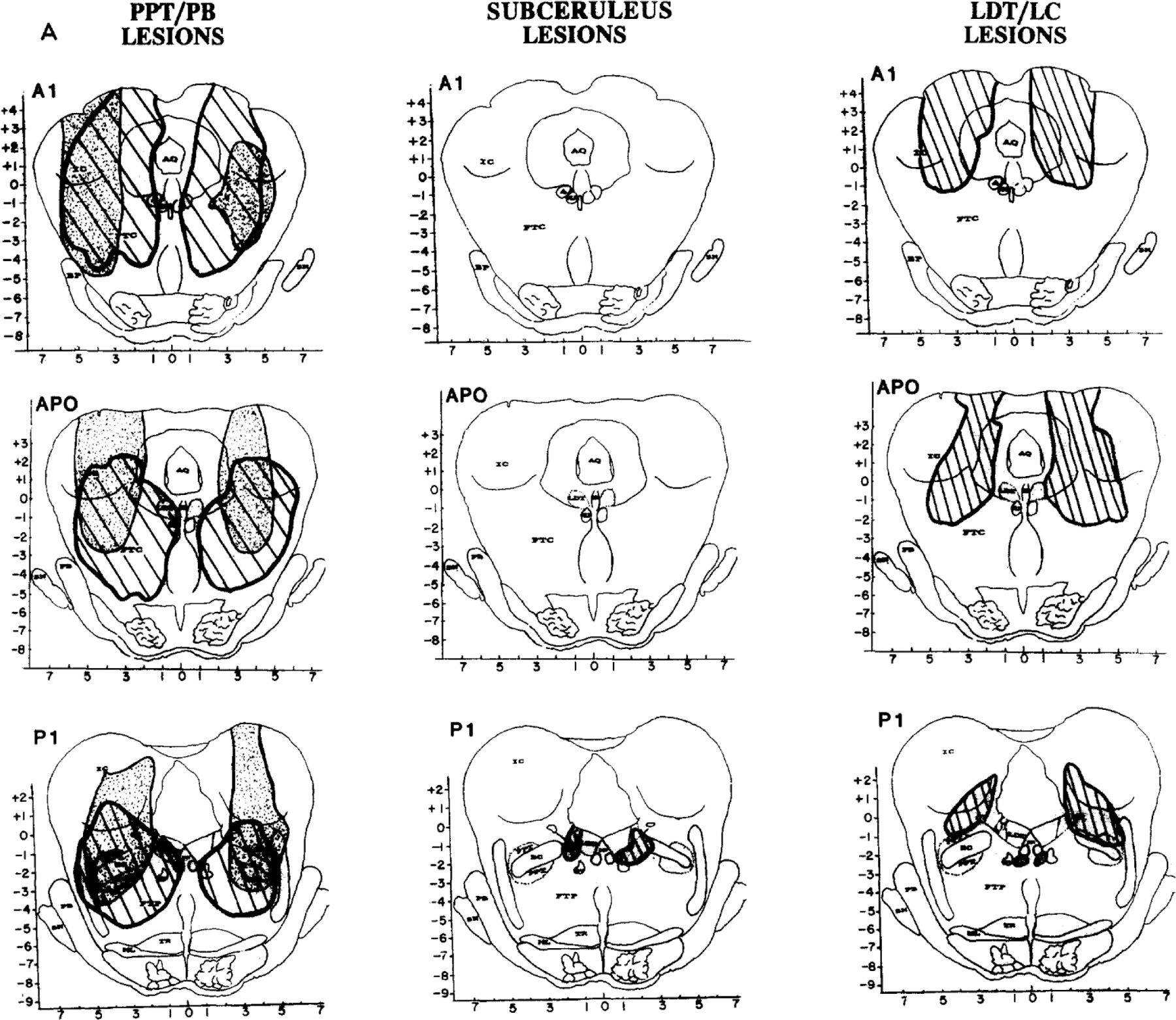
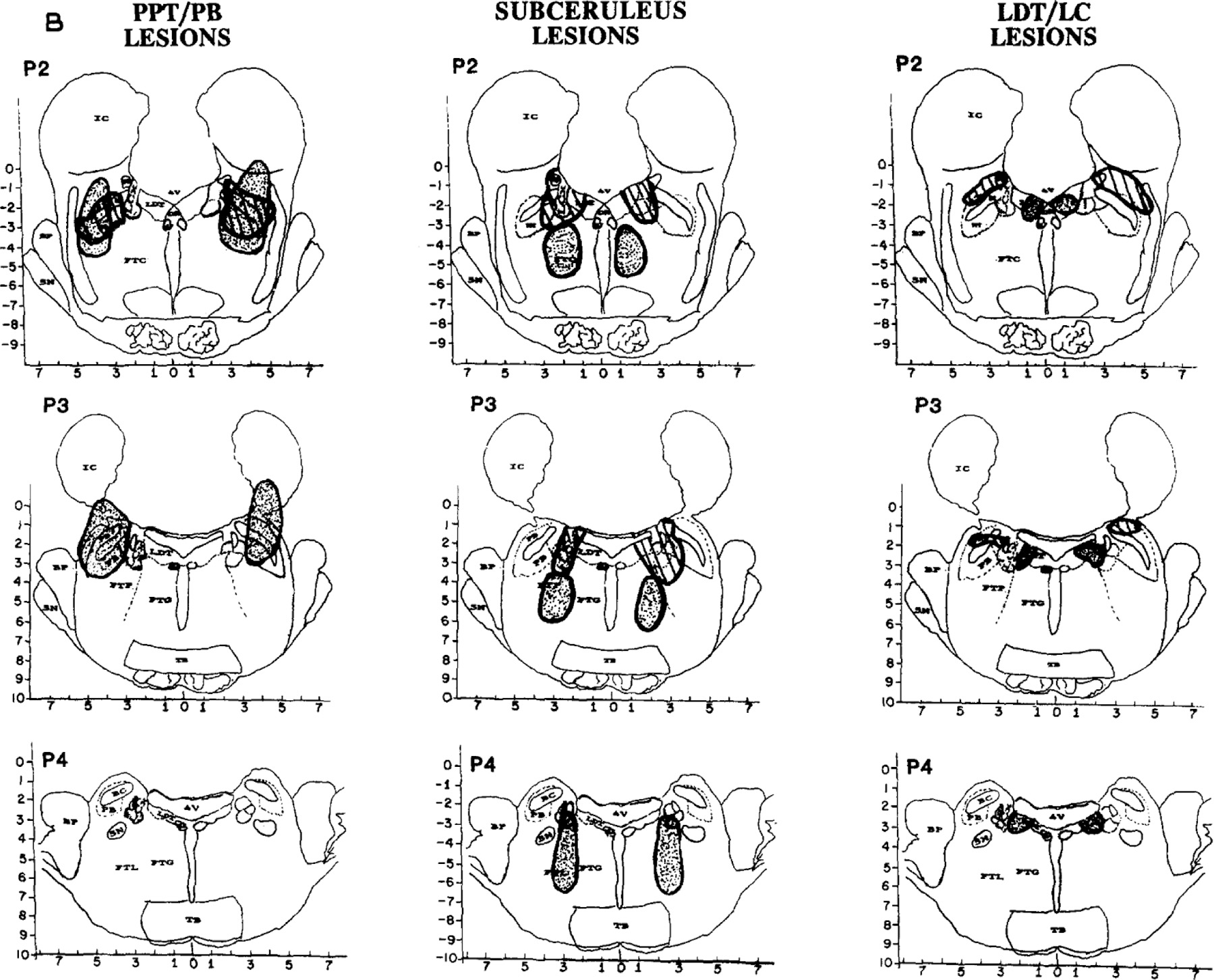
B. Reconstructions of lesions in 2 cats (one dotted and the other striped) in each of the 3 lesion groups, PPT/PB (left), subceruleus (middle) and LDT/LC (right). Coronal section and abbreviations are adapted from the Berman atlas and approximate 1 mm intervals from Al throught P4. Left panel: Group 1 PPT/PB. Lesions were larger than in the other 2 groups and involved most of the lateral pontine cholinergic cell columns, including PFT and PB, often encompassed the central tegmental field (FTC) and sometimes the parvocellular tegmental field (FTP) as well. Tissue lost posterior to P2 is illustrated in 1 cat (dotted lesion); damage to the peribrachial (PB) cholinergic columns is evident at P3. Middle panel: Group 2 subceruleus. Lesions were smaller as well as caudal and ventromedial to PPT/PB lesions. Peri-LC alpha was damaged in all 6 cats. Some lesions encompassed locus ceruleus complex (LCx; striped lesion), and others damaged peri-LC alpha together with the underlying reticular formation (dotted), mostly the lateral tegemental field (FTL) and some of the gigantocellular tegmental field (FTG). Right panel: Group 3 LDT/LC. One cat in this group had a large lateral lesion involving most of FTC and some of the LC (striped). The other lesions in the group were more medial (dotted), encompassing all or parts of the lateral dorsal tegmenturn (LDT) and/or locus ceruleus (LC).