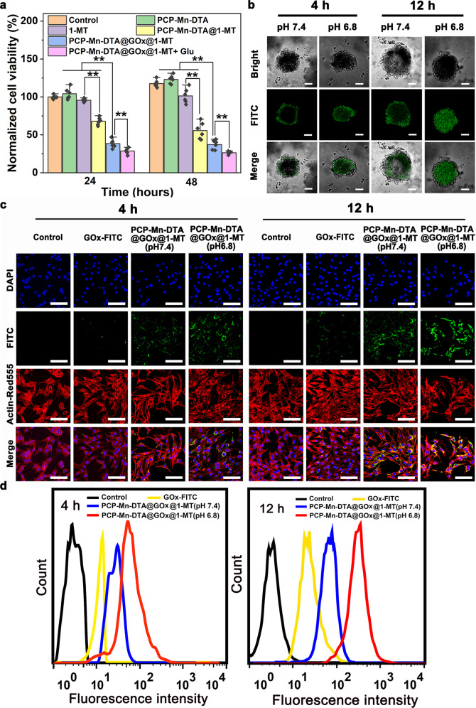
Fig. 5. In vitro cytotoxicity, cellular uptake, and tumor penetration studies of PCP-Mn-DTA@GOx@1-MT nanosystem.
a Cytotoxicity studies of B16F10 cells cultivating with PCP-Mn-DTA, 1-MT, PCP-Mn-DTA@1-MT, and PCP-Mn-DTA@GOx@1-MT with or without glucose for 24 h and 48 h, respectively. b Tumor penetration and (c) endocytosis images of PCP-Mn-DTA@GOx@1-MT labelled with FITC in B16F10 MCSs after respective incubation at pH 6.8 or 7.4 for 4 h and 12 h, with images representative of 3 experiments. Nuclei and cytoskeleton were respectively stained with DAPI (blue) and ActinRed™555 (red). d Quantitative FCM analysis based on (c). Data represent mean ± SD (n = 6 biologically independent samples). P-values were determined by unpaired Student’s t-test (two-tailed), **p < 0.01. Scale bars: 100 µm for b, 50 µm for c.

