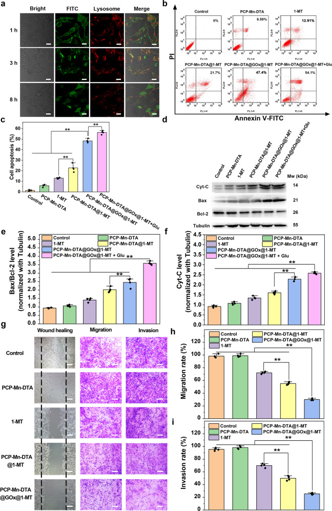
Fig. 7. In vitro lysosomal escape, cell apoptosis and anti-metastasis studies of PCP-Mn-DTA@GOx@1-MT nanosystem.
a B16F10 cells incubated with FITC-labelled PCP-Mn-DTA@GOx@1-MT for different times, followed by staining with Lysotracker red to image the lysosome escape, with images representative of 3 experiments. b, c FCM and quantitative apoptosis analysis of B16F10 cells after difference treatments for 24 h. d–f Apoptosis-associated proteins in B16F10 cells after the treatment with indicated systems examined by western blot (d), and the related quantitative analysis of Bax/Bcl-2 (e) and Cytochrome C (f). g–i Microscopy images (g) and quantitative analysis of the wound healing and migration (h) as well as invasion assays (i) of B16F10 cells after the treatment with these systems. Data represent mean ± SD (n = 4 biologically independent samples). P-values were determined by unpaired Student’s t-test (two-tailed), **p < 0.01. Scale bars: 20 μm for a, 100 μm for g.

