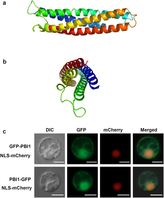
Fig. 3. PBI1, with a four-helix bundle structure, localizes mainly to the nucleus.
a Side view of PBI1, which forms a four-helix bundle. Coloring is from blue at the N-terminus to red at the C-terminus. b End view, with N- and C-termini at the front. c Detection of GFP-PBI1 and PBI1-GFP after transient expression in rice protoplasts. mCherry with a nuclear localization signal was used as a nuclear localization marker. Scale bar = 10 µm. Similar results were obtained in three independent experiments.

