Figure 1.
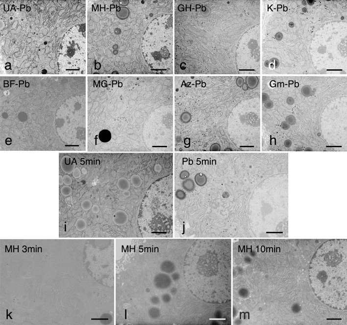
(a–h) EM images of mouse liver stained with various dyes followed by RPb. Ultrathin sections of mouse liver fixed using the conventional 2% glutaraldehyde and 1% osmium tetroxide fixation and embedded in epoxy resin, doubly stained with (a) UA and RPb (UA–Pb), (b) MH and RPb (MH–Pb), (c) Gill No.3 hematoxylin and RPb (GH–Pb), (d) Kernechtrot and RPb (K–Pb), (e) basic fuchsin and RPb (BF–Pb), (f) methyl green and RPb (MG–Pb), (g) alizarin and RPb (Az–Pb), and (h) Giemsa solution and RPb (Gm–Pb). Hepatocytes, including the nucleus and cytoplasmic organelles, were stained clearly to different degrees of contrast with all dyes upon post-staining with RPb. The contrast of almost all organelles was highly enhanced upon double staining with MH and RPb, to the same extent as that with UA and RPb. Bar = 2 μm. (i—m) Ultrathin sections of mouse livers solely stained with (i) UA for 5 min, (j) RPb for 5 min, (k) MH for 3 min, (l) MH for 5 min, and (m) MH for 10 min. Cells were not clearly displayed in the 3 or 5 min MH single staining, but slight contrast enhancements were observed in the U, RPb and 10 min MH stainings. Bar = 2 μm. The results represent at least three independent experiments with similar results.

