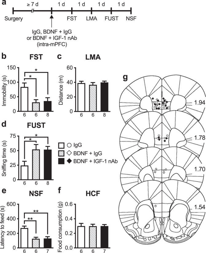
Fig. 4. The antidepressant-like effects of intra-mPFC infusion of BDNF are not blocked by co-infusion of IGF-1 nAb.
a Experimental timeline for behavioral testing after intra-mPFC infusion of control IgG (160 ng/side), BDNF (100 ng/side) plus control IgG, or BDNF plus IGF-1 nAb (160 ng/side). b Immobility time in the forced swim test (FST) 1 day after intra-mPFC infusion (F2,17 = 7.45, p = 0.0111, n = 6–8). c Locomotor activity (LMA) 2 days after intra-mPFC infusion (F2,17 = 0.514, p = 0.607, n = 6–8). d Time spent sniffing female urine in the female urine sniffing test (FUST) 3 days after intra-mPFC infusion (F2,17 = 5.14, p = 0.0179, n = 6–8). e Latency to feed in the novelty-suppressed feeding (NSF) test 4 days after intra-mPFC infusion (F2,16 = 12.4, p = 0.0006, n = 6–7). A BDNF + IGF-1 nAb-infused mouse was not subjected to the NSF because the mouse became lethargy, and exhibited ruffled fur and hunched posture as a result of overnight food deprivation. f Home cage feeding (HCF) just after the NSF (F2,16 = 2.98 × 10⁻¹⁵, p > 0.999, n = 6–7). g Schematic representation of mPFC infusion sites. Plates are from ref. [50]; 1.94, 1.78, 1.70, and 1.54 indicate distances (mm) from bregma. Data are expressed as means ± SEM. *p < 0.05, **p < 0.01 (one-way ANOVA followed by Tukey’s post hoc test).

