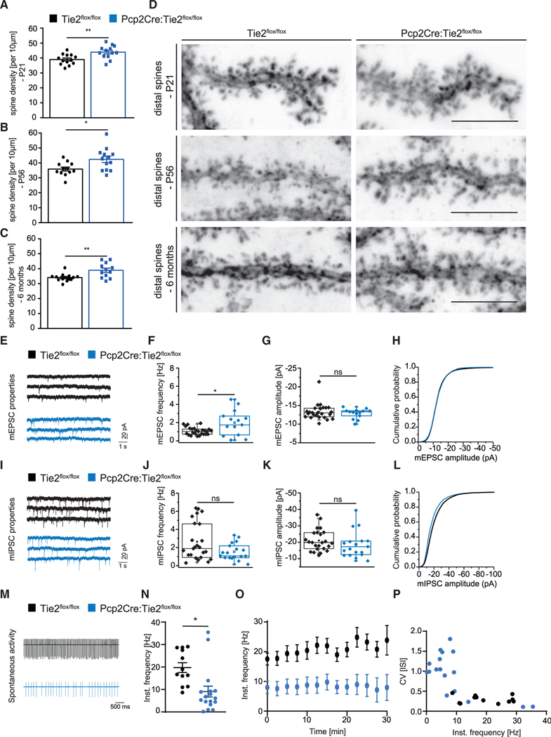
Figure 6. Loss of Tie2 expression in postnatal PCs results in alterations of cerebellum network functionality.
(A–D) Distal dendritic spines were counted in AAV8-transduce PCs of Tie2flox/flox and Pcp2Cre:Tie2flox/flox animals at P21 (A and D), P56 (B and D), and 6 months (C and D). Data are represented as mean ± SEM from a minimum of n = 10 neurons from a minimum of three independent animals. Unpaired Student’s t test; *p < 0.05; **p < 0.01. Representative pictures are shown in (D). Scale bar: 5 μm.
(E–L) PCs of Tie2lox/lox and Pcp2Cre:Tie2flox/flox animals were patched at 8–11 weeks of age and mEPSCs (E–H) and mIPSCs (I–L) were recorded. Example traces are shown in (E) and (I). Event frequency (F and J), amplitude (G and K), and cumulative probability (H and L) are shown. Data are represented as boxplot from a minimum of n = 14 neurons from a minimum of 4 independent animals.
(M–P) PCs of Tie2lox/lox and Pcp2Cre:Tie2flox/flox animals were patched at 8–11 weeks of age and spontaneous tonic firing was recorded. Example traces are shown in (M). Instantaneous frequency (N–P) was analyzed. Data are represented as mean ± SEM from a minimum of n = 12 neurons from a minimum of 3 independent animals. Unpaired Student’s t test; *p < 0.05; ns, not significant. See also Figure S4.

