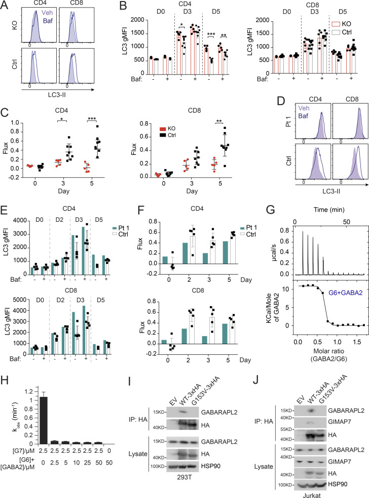
Figure 3.
Defective autophagy in T cells from Pt 1 and Gimap6−/− mice. (A) Whole splenocytes from Gimap6−/− (KO) or Gimap6+/+ (WT control, Ctrl) mice were treated with 1 μg/ml each of anti-CD3/CD28 for 3 d and then with 10 nM Baf or vehicle (Veh) for 2 h. Flow cytometry of LC3 expression (LC3-II) in CD4+ and CD8+ T cells. (B) Quantification of A for the indicated days. **, P < 0.01 (unpaired Student’s t test). nKO = 5; ncontrol = 8. (C) Autophagic flux of A, calculated as (gMFI LC3Baf − gMFI LC3Veh)/gMFI LC3Veh. *, P < 0.05; **, P < 0.01; ***, P < 0.001 (unpaired Student’s t test). nKO = 5; ncontrol = 8. Shown is one of two independent repeats. gMFI, geometric MFI. (D) Flow cytometry histograms of LC3-II in human PBMCs from Pt 1 and healthy controls (Ctrl) stimulated with 1 μg/ml each of anti-CD3/CD28 for 3 d before treatment with Baf or vehicle (Veh) followed by flow cytometry and gating on CD4+ and CD8+ cells. (E) Quantification of gMFI of LC3-II of D as in B for the indicated days. (F) Quantification of autophagic flux of D as in C. Shown is one of two independent repeats and each dot indicates an individual donor in E and F. (G) 600 µM GABARAPL2 (GABA2) was titrated into 50 µM GIMAP6 (R134D; G6) and the resulting heat change was monitored in an ITC device. Data were fitted to a KD of 40 + −10 nM (n = 0.63 + −0.01). Shown is one of three experiments. (H) GTPase inhibition of 2.5 μM GIMAP7 (G7) by various concentrations of a 1:1 M GIMAP6 (G6)-GABARAPL2 (GABA2) complex. Shown is one of three experiments. (I) WB of HEK293T cells transfected with empty vector (EV), HA-tagged GIMAP6WT (WT-HA), or HA-tagged GIMAP6G153V (G153V-HA) for 1 d. GIMAP6WT and GIMAP6G153V were immunoprecipitated with HA tag antibody. The cell lysate and IP were probed for GABARAPL2, HA, and HSP90. Shown is one of three experiments. (J) WB of Jurkat cells transduced with lentivirus of empty vector (EV), HA-tagged GIMAP6WT (WT-HA), or HA-tagged GIMAP6G153V (G153V-HA) and selected with puromycin (1 µg/ml) for 4 d. GIMAP6WT and GIMAP6G153V were immunoprecipitated with HA tag antibody. The cell lysate and IP were probed for GABARAPL2, GIMAP7, HA, and HSP90. Shown is one of three experiments. Bars (B, C, E, F, and H) represent mean ± SEM.

