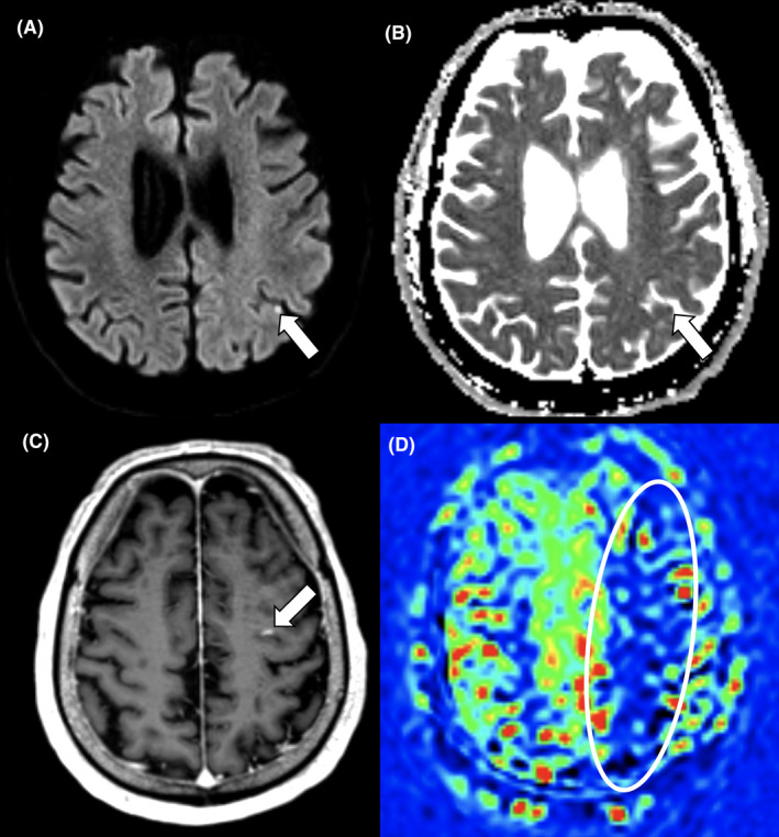
FIGURE 1.

Cortical microinfarcts as an incidental imaging finding. Brain MRI at 3‐month follow‐up in a 68‐year‐old male COVID‐19 critical care survivor revealing four asymptomatic cortical microinfarcts of varying age and a hypoperfusion syndrome in the watershed areas of the left middle cerebral artery territory. Diffusion‐weighted b1000 image (A) and Apparent Diffusion Coefficient map (B) revealing an acute cortical microinfarct in the left parietal lobe. Subacute cortical microinfarct with blood–brain barrier disruption revealed on a contrast‐enhanced 3D T1‐weighted image (C). Reduced relative cerebral blood flow in the watershed areas of the left middle cerebral artery territory detected by Arterial Spin Labeling (D)
