
Figure 2.
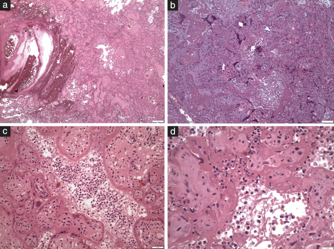
Hematoxylin‐and‐eosin‐stained placental sections showing histopathology of SARS‐CoV‐2 placentitis: (a) fresh intervillous thrombosis (×45); (b) massive perivillous fibrinoid deposition (×100); (c) inflammation in the intervillous space (intervillositis) and damage of the perivillous trophoblast (×200); and (d) inflammatory cells of mixed type within a meshwork of fibers in the intervillous space (×400).