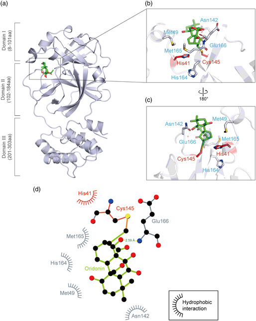
Figure 5.

Crystal structure of SARS‐CoV‐2 3CLpro in complex with Oridonin. a) Overview of the structure of Oridonin‐bound 3CLpro. The protein is shown as a cartoon in blue‐white and Oridonin is presented as green sticks. b–c) Zoomed‐in view of the binding site of Oridonin. The catalytic residues His41 and Cys145 are shown in red sticks, and others residues interacting with Oridonin are shown in blue‐white sticks. The hydrogen bonds formed with the residues of 3CLpro are indicated as yellow dashed lines. d) 2D presentation of Oridonin binding. The ligand Oridonin and the residues forming polar interactions are shown in ball‐and‐stick representations, the C—S covalent bond is shown in orange, the hydrogen bond is shown as a yellow line with the distance labeled, and other residues contribute to hydrophobic interactions.
