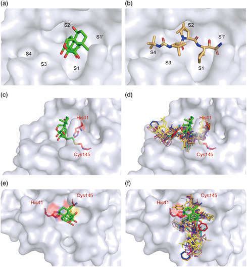
Figure 6.

Binding mode of Oridonin with 3CLpro. a) Oridonin only occupies the S1 of the substrate‐binding pocket. b) Boceprevir occupies the S1, S2, S3, and S4 subsites of the substrate‐binding pocket. c) The structure of 3CLpro is shown in surface representation in blue‐white, Oridonin is shown as green stick, and the two catalytic residues are shown as red sticks. d) The known structures of 3CLpro bound to inhibitors are superimposed on the 3CLpro‐Oridonin complex. Different colors represent reported inhibitors, which were shown as lines (PDB codes: 7D1M, 7D10, 7C8T, 7C7P, 7C8R, 7COM, 7JKV, 6W63, 5RGY, 5RH5, 6Y2F, 6XHM, 7BRP, 5RH6, and 5RH7). e,f) The top views of (c) and (d), respectively.
