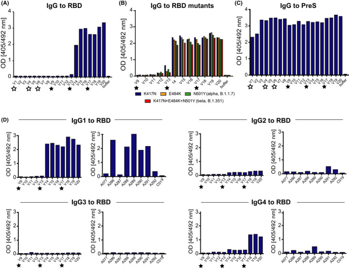
FIGURE 4.

Development of specific antibody responses in the immunized subject. Serum IgG reactivity to (A) folded RBD after immunization with unfolded E. coli (white stars) and folded HEK cell‐expressed (black stars) PreS‐RBD (x‐axes, time points). (B) IgG reactivity to RBD mutations K417N, E484K, N501Y (alpha, B.1.1.7), and K417N+E484K+N501Y (beta, B.1.351) after immunization with HEK cell‐expressed PreS‐RBD at different time points (x‐axis). (C) PreS‐specific IgG after immunization with unfolded E. coli (white stars) and folded HEK cell‐expressed (black stars) PreS‐RBD (x‐axes, time points) and (D) IgG subclass analysis of immunization with HEK cell‐expressed PreS‐RBD (left) at different time points (x‐axes) and of subjects (x‐axes) 4 weeks after full immunization with licensed COVID‐19 vaccines (right). Sera were diluted 1:50; OD values are average values of duplicate determinations with <5% deviation (y‐axes) and correspond to amounts of bound antibodies
