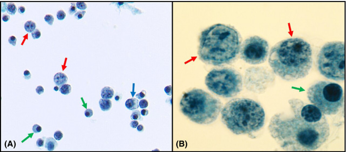
FIGURE 1.

(A) BAL ThinPrep showing immunoblasts (red arrows), plasmacytoid lymphocytes (blue arrows), and mature plasma cells (green arrows) (Papanicolaou staining, magnification ×40). (B) Oil immersion image of BAL showing immunoblasts (red arrows) and plasma cells (green arrow) (Papanicolaou staining, magnification ×100)
