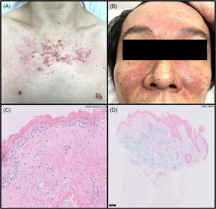
FIGURE 1.

(A) and (B) Erythematous rash with telangiectasis on chest and both medial cheeks sparing nasolabial folds. (C) Histology from the punch biopsy of the chest lesion shows epidermal atrophy, vacuolar interphase changes with sparse inflammatory infiltrate. (D) Mucin, highlighted by special stain Alcian blue, is noted in the superficial dermis
