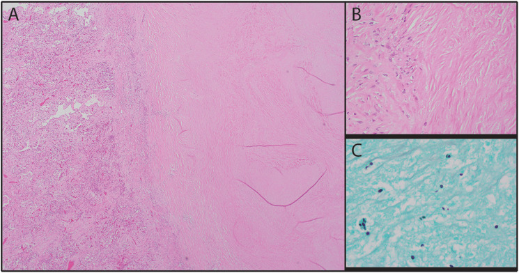
Figure 3.
Subpleural necrotizing granuloma. A-B: The lesion consisted of a necrotic core surrounded by epithelioid histiocytes (A: H&E, × 40; B: H&E, × 400). C: The necrotic core contained numerous yeast forms with narrow-based budding consistent with Histoplasma (Grocott methenamine silver, × 1000).

