Fig. 5.
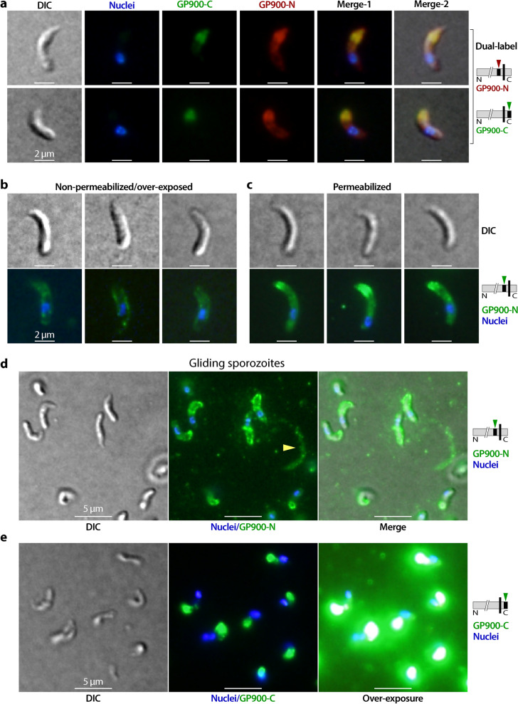
IFA detection of GP900 protein in excysted sporozoites. a Dual-labeling of GP900 with mouse anti-GP900-N (red) and rabbit anti-GP900-C (green) in excysted sporozoites. b, c IFA detection of GP900 protein on the external surface of sporozoites that had been fixed in formaldehyde but were unpermeabilized (b) in comparison with permeabilized sporozoites (c), using anti-GP900-N antibody. To facilitate a good comparison, both samples were processed in parallel under the same experimental conditions. It should be noted that weak, but clear when over-exposed, signals were produced in unpermeabilized sporozoites. d, e IFA detection of GP900 protein on the trails of gliding sporozoites using anti-GP900-N (d) and anti-GP900-C (e) antibodies; anti-GP900-N antibody stained the surface of sporozoites and some gliding trails (d) while anti-GP900-C antibody only stained the apical region of the gliding sporozoites (e). In all panels, nuclei were stained with DAPI

