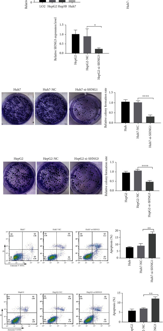
Figure 11.

SHNG1 and SHNG3 inhibit apoptosis and promote proliferation of HCC cells. (a) Expression of SHNG1 in LO2, HepG2, Hep3B, and Huh7 cell lines was measured by qRT-PCR. (b) siRNA knockdown efficiency was confirmed by qRT-PCR in Hun7 cells. Huh7: Huh7 cells without transfection; Huh7-NC: Huh7 cells transfected with negative control (NC); Huh7-si-SHNG1: Huh7 cells transfected with si-SHNG1. (c) siRNA knockdown efficiency was confirmed by qRT-PCR in HepG2 cells. HepG2: HepG2 cells without transfection; HepG2-NC: HepG2 cells transfected with negative control; HepG2-si-SHNG3: Huh7 cells transfected with si-SHNG3. (d, e) Colony formation assay was used to assess the effect of siSHNG1 in Huh7 cells and the effect of siSHNG3 in HepG2 cells. (f, g) Flow cytometry analysis of Annexin V-FITC/PI staining was used to assess the number of apoptotic cells in each treatment in Huh7 and HepG2 cells. ∗p < 0.05; ∗∗p < 0.01; ∗∗∗p < 0.0001.
