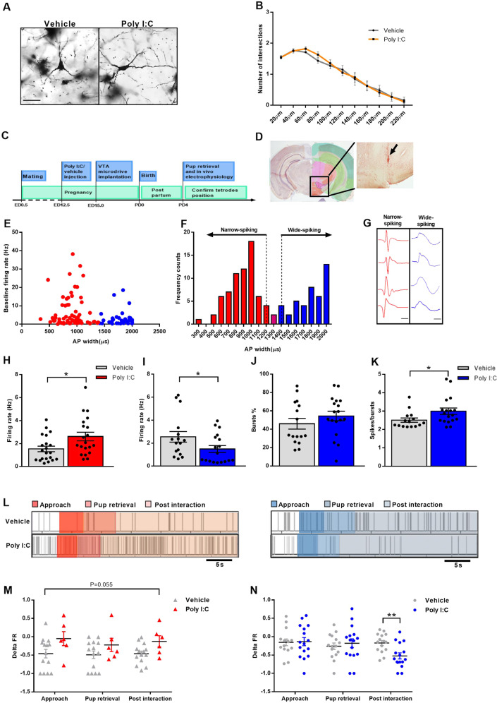
Fig. 5. VTA single units show altered firing rate (FR) during baseline and pup exposure.
A Examples of Golgi–Cox impregnated VTA neurons from vehicle and Poly I:C-injected mothers (20X magnification; scale bar: 50 μm). B Poly I:C injection does not result in alterations of number of intersections in VTA neurons of mothers (N = 5 animals/group). The morphology of six neurons was reconstructed from two sections per animal for a total of 30 neurons per group. C Schematic depiction of the experimental timeline for in vivo electrophysiology. D Representative coronal section from a control mother showing the tetrode tract mark in the VTA region (arrow) (adapted from Allen Brain Atlas; Bregma: −2.555). E Baseline FR versus full AP widths of recorded single units (N = 111 neurons; five animals/group; red: narrow-spiking neurons; blue: wide-spiking neurons). F Frequency histogram of full AP widths showing a bimodal distribution. Cutoffs of 1200 µs and 1400 µs spike width were used to differentiate between two subpopulations of neurons (narrow-(red) and wide-(blue) spiking neurons) within the VTA (arrows). Single units with spike width comprised between 1200 µs and 1400 µs were assigned to either group based on defined waveform characteristics or excluded from the analysis (N = 111 neurons; 5 animals/group). G Examples of single units’ waveforms from narrow-(red) and wide-(blue) spiking neurons (scale bar: 500 µs). H, I Poly I:C injection during pregnancy leads to H significant FR increase of narrow-spiking neurons (U = 122; Ρ = 0.022; Ν = 20–21 neurons/group, five animals/group) and I significant FR reduction of wide-spiking neurons (U = 86; Ρ = 0.046; Ν = 16–18 neurons/group, five animals/group). J, K Poly I:C-injected mothers J do not show any change in bursts percentage (%) of wide-spiking neurons (N = 16–18 neurons/group; five animals/group) but (K) have significantly altered number of spikes/bursts (U = 75.5; Ρ = 0.03; Ν = 16–18 neurons/group; five animals/group). *Ρ < 0.05. L Sample raster plots of narrow- and wide-spiking neurons during retrieval of the last pup in MIA and control mothers; behavioral epochs are marked in different shades of colors (from left to right: approach, pup retrieval and post interaction. Narrow-spiking neurons: shades of red; wide-spiking neurons: shades of blue). M Narrow-spiking neurons show a trend for alteration in FR change during the three behavioral epochs in Poly I:C-injected mothers. Ρ value indicates the main effect of treatment (main effect of treatment: F([1) = 4.230; Ρ = 0.055; Ν = 6–14 neurons/group; 3–4 animals/group). N Wide-spiking neurons show altered FR change after Poly I:C injection during the post interaction behavioral epoch. Asterisks refer to the pairwise comparison. Delta FR: calculated according to the standardized change ratio described in “Materials and methods” (interaction of treatment and behavior: F(2) = 4.954; Ρ = 0.01, Ν = 15–16 neurons/group; 3–4 animals/group). All data are presented as mean ± SEM, *Ρ < 0.05, **Ρ < 0.01.

