
Figure 2.
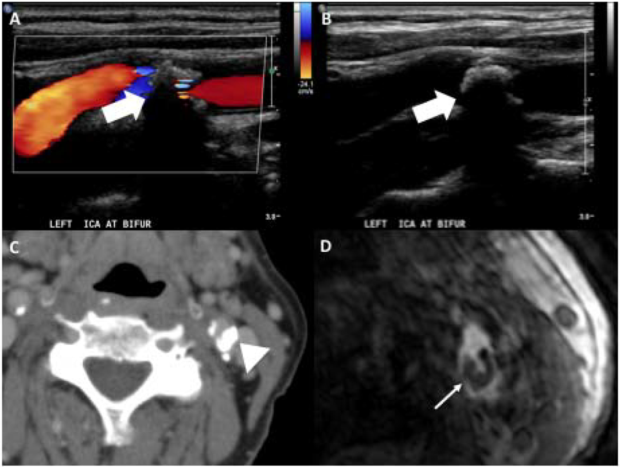
85-year-old female with large calcified plaque at the left carotid bifurcation as seen on US, CT, and MR. Doppler (A) and gray scale (B) US images demonstrate large echogenic plaque with posterior acoustic shadowing (white large arrows). The posterior acoustic shadowing limits appreciation of the size of the plaque. CT on the same patient (C) shows large calcified plaque in the posterior aspect of the left carotid bifurcation (arrowhead). Axial slice of 3D MR TOF (D) shows area of hypointensity in the posterior aspect of the left carotid bifurcation corresponding to the plaque calcification (narrow white arrow).