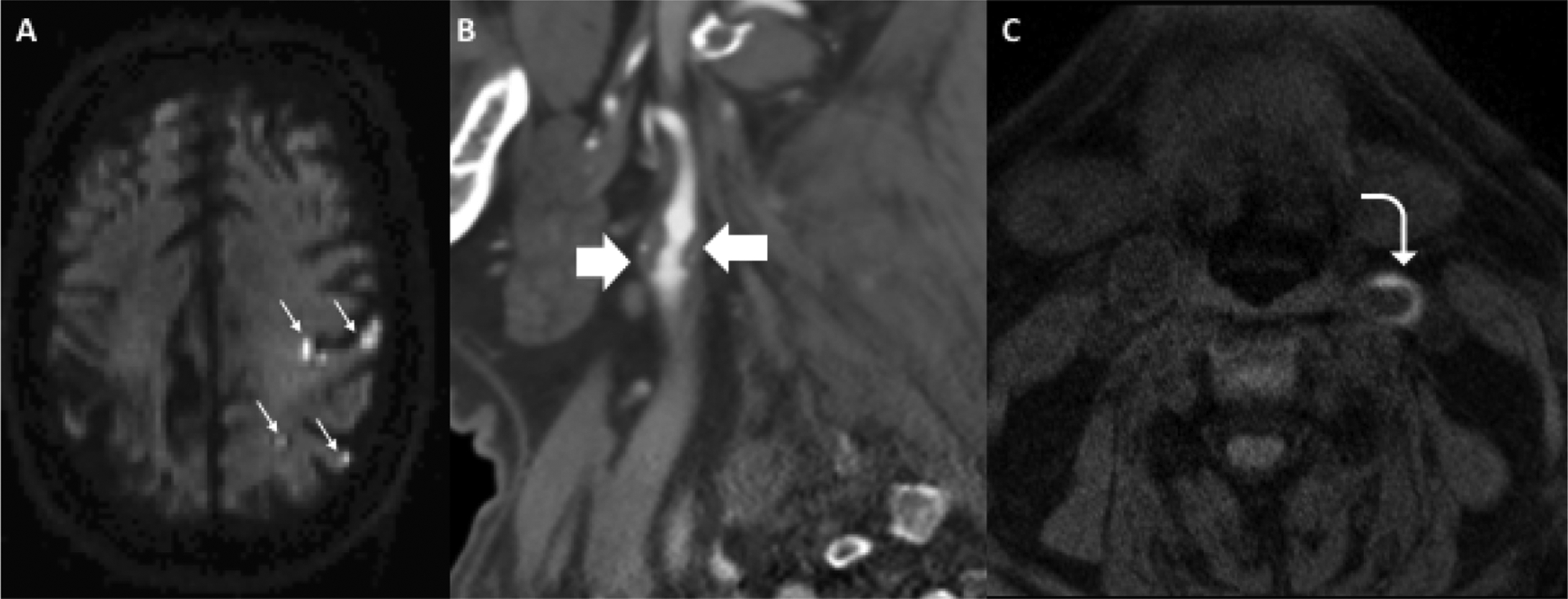
Figure 5.

Case study of 71-year-old female presenting with acute right-sided weakness found to have multiple acute infarctions throughout the left cerebral hemisphere on axial DWI (A, small white arrows). Immediately after rapid brain MRI, she had a CTA head and neck which showed a large soft/fibrofatty plaque in the proximal left ICA (B, block arrows) with areas of ulceration resulting in less than 50% stenosis by NASCET criteria. On axial MPRAGE, she had areas of crescentic T1 hyperintensity (C, curved arrow) consistent with intraplaque hemorrhage.
