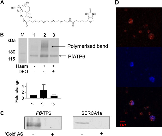
FIG 2.
Artemisinins’ interaction with PfATP6. (A) DHA-biotin probe structure. (B) Western blots of PfATP6-enriched microsome pulldowns that were untreated (lane 1), preincubated with 200 μM heme (lane 2), or preincubated with heme and 200 μM DFO, which is an iron chelator (lane 3). Fold change refers to the band intensity relative to untreated PfATP6 (lane 1), calculated from densitometry. PfATP6-enriched microsomes were incubated with DHA-biotin before the addition of streptavidin-coated magnetic beads (Invitrogen, UK), which were pulled down using a magnetic rack. Beads were released by incubation in SDS at 95°C for 5 min. The supernatant was sampled for SDS-PAGE. (C) Western blots of PfATP6- and SERCA1a-enriched microsome pulldowns preincubated with artesunate (AS) before pulldowns were performed with DHA-biotin. (D) Immunofluorescence assay with P. falciparum parasites within red blood cells. Trophozoite-stage parasites were preincubated with DHA-biotin before staining with 1 μg/mL tetramethyl rhodamine isocyanate (TRITC)-tagged antibiotin antibody (red) (top) and the nucleic acid-specific stain DAPI (blue) (middle). (Bottom) Merge.

