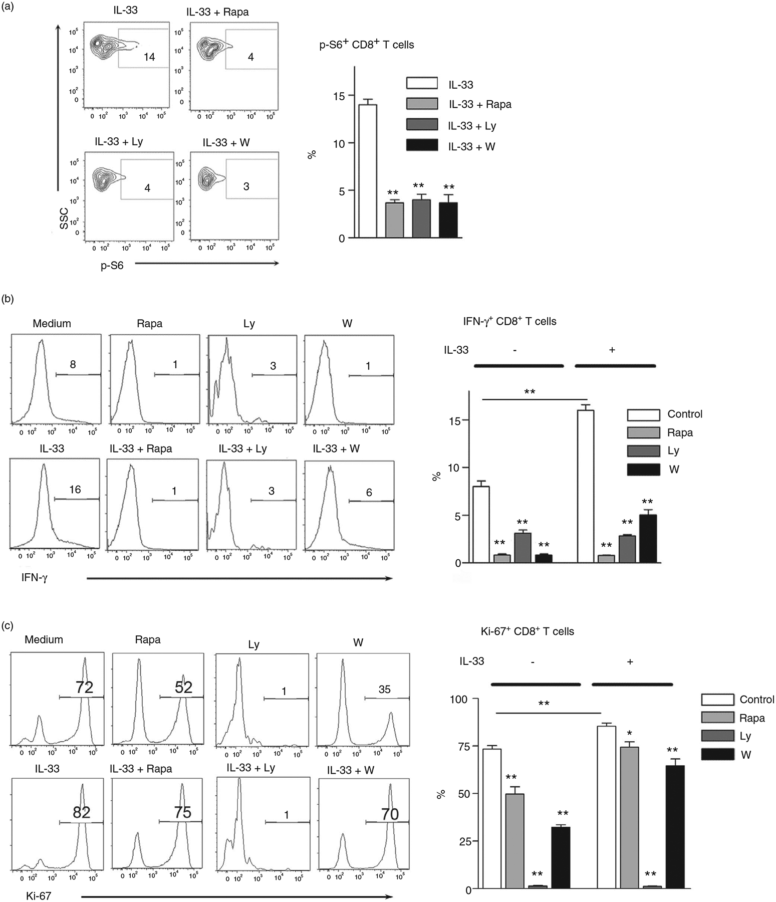
FIGURE 3.

Inhibition of mTORC1 results in impaired CD8 T-cell activation and proliferation by IL-33. (a) Splenocytes isolated from naïve mice were cultured in anti-CD3/CD28-coated plates for 3 days. Cells were treated with rapamycin (25 nM), Ly294002 (5 μM) and wortmannin (100 nM) for 2 h, followed by 100 ng/ml IL-33 stimulation for 10 min at 37°C. DMSO was used as a control. The p-S6 expression was evaluated according to the BD Phosflow staining protocol, and the percentages of p-S6+ cells were shown. The IL-33 group was used as a control for comparison (b and c) Purified CD8+ T cells were cultured in anti-CD3/CD28-coated plates in the presence of IL-33 (100 ng/ml) and inhibitors for 4 days. Brefeldin A was added in the last 6 h of culture. Intracellular IFN-γ and Ki-67 expression were analysed by flow cytometry. The data are shown as mean ± SEM from single experiments and are representative of at least two experiments performed. Triplicates were performed for each group. One-way ANOVA with Dunnett multiple comparisons was used for statistical analysis. *p < 0·05; **p < 0·01
