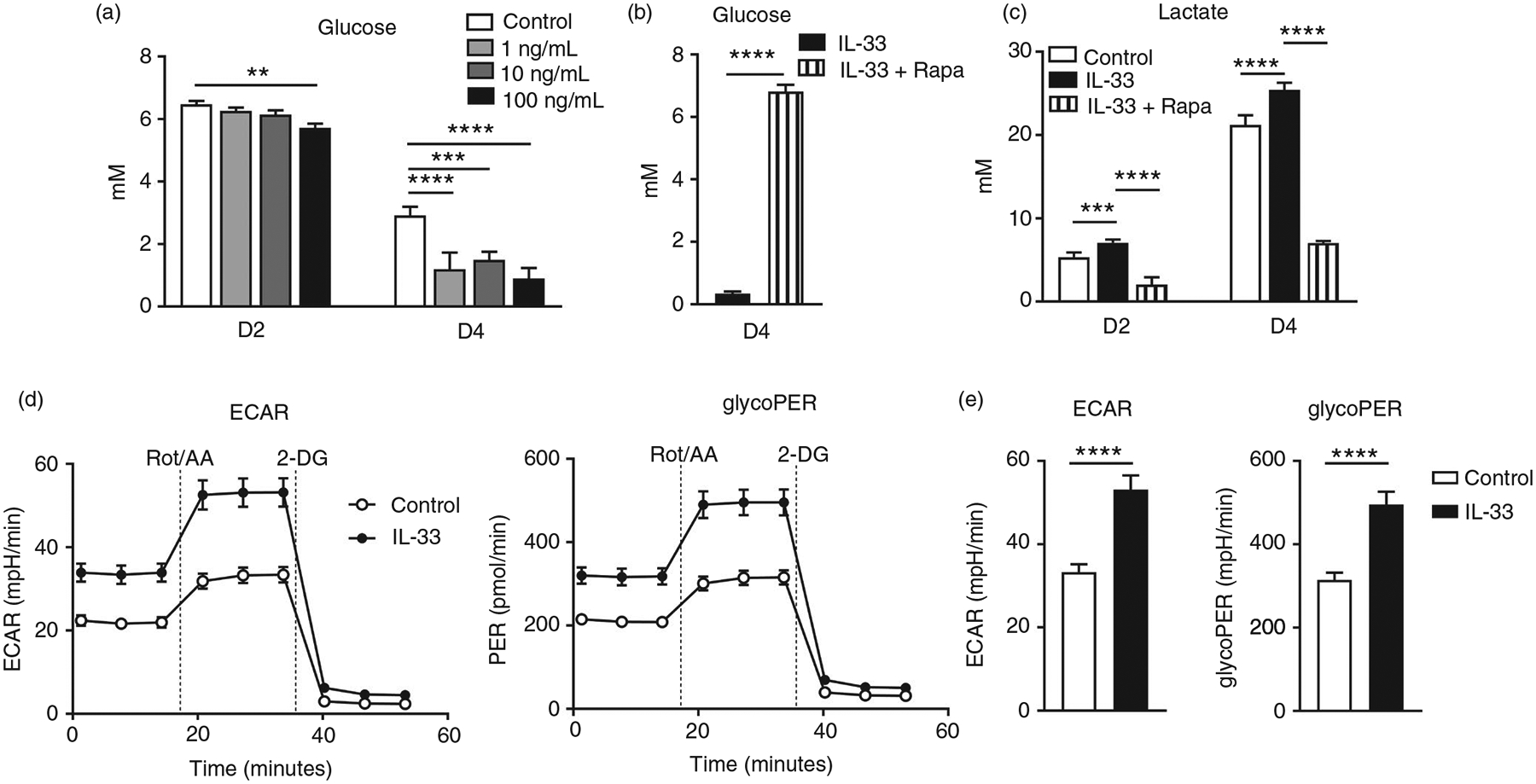
FIGURE 4.

IL-33 promotes glucose uptake and lactate production in CD8 effector cells. CD8+ T cells were purified from naïve mouse spleens and cultured in anti-CD3/CD28-coated plates in the presence of different doses of IL-33. (a) Supernatant was collected at days 2 and 4 for the measurement of glucose. (b and c) CD8+ T cells were cultured in the presence of IL-33 (100 ng/ml) with rapamycin (25 nM) added or omitted. Supernatant was collected for glucose and lactate measurement. (d–e) WT CD8+ T cells were cultured in anti-CD3/CD28-coated plates with or without IL-33 (100 ng/ml) for 3 days. Cells were harvested and seeded into a 96-well Seahorse plate at a density of 1 × 10⁵/well in Seahorse XF assay medium. Glycolytic rate assay kit (Agilent, Santa Clara, CA) was used to determine extracellular acidification rate (ECAR) and glycolytic proton efflux rate (glycoPER) according to the manufacturer’s instruction. The data are shown as mean ± SEM from single experiments and are representative of at least two experiments performed. Triplicates were performed for each group. A two-tailed Student’s t test was used for statistical analysis of two groups. One-way ANOVA with Dunnett multiple comparisons were used for statistical analysis of three or more groups. **p < 0·01; ***p < 0·001; ****p < 0·0001
