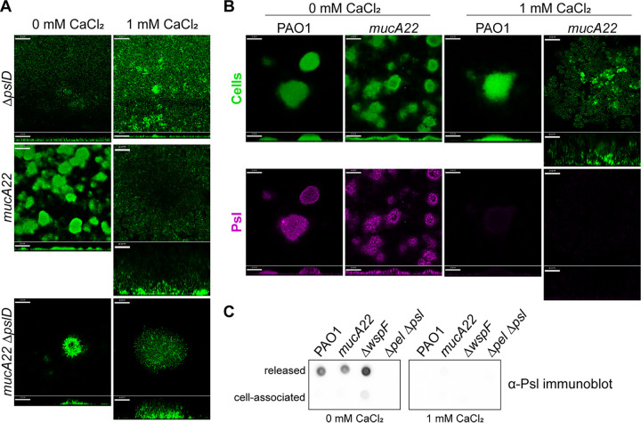
FIG 4.
Psl is not required for the formation of calcium-cross-linked mucoid biofilms. (A) Biofilm growth of PAO1 strains deficient in Psl production (ΔpslD). Representative confocal images are from biofilms cultivated for 72 h under continuous-flow conditions. Cells are constitutively expressing GFP (pseudocolored green). Magnification, ×200. Bars, 50 μm. (B) Psl localization in nonmucoid and mucoid 72-h-old flow cell biofilms grown with and without calcium. Cells are constitutively expressing GFP (pseudocolored green). Psl was visualized using a fluorescent Psl-specific lectin (HHL-TRITC) (magenta). Horizontal cross sections (squares) from the middle of the biomass and sagittal views (rectangles) are shown. Magnification, ×200. Bars, 50 μm. (C) Anti-Psl immunoblotting performed on released and cell-associated fractions of statically grown biofilms. Cultures were grown for 24 h at 37°C in 5 mL of EPRI medium in wells of a 6-well tissue culture plate.

