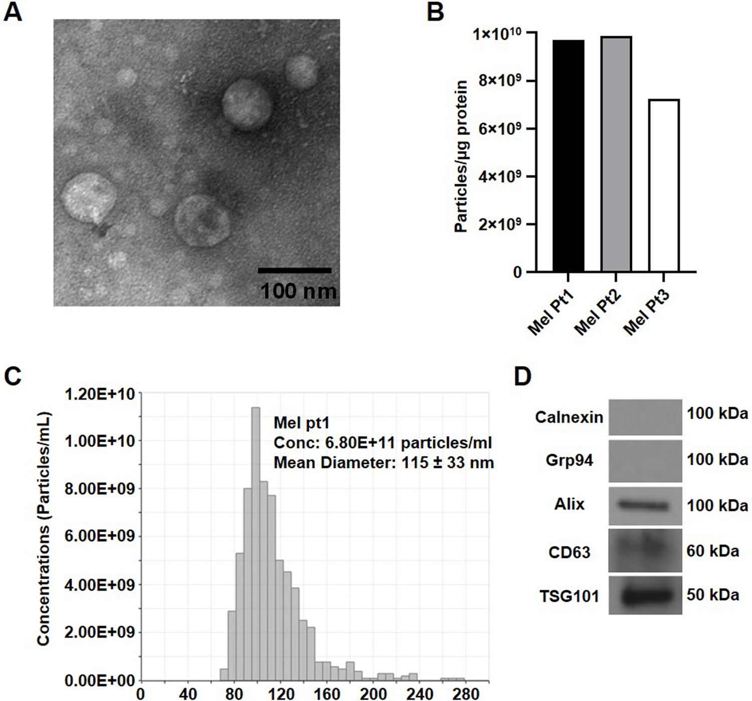
Fig. 2:

Characterization of exosomes isolated using mini-SEC from the plasma of melanoma cancer patients. (A) TEM images of exosomes recovered in the SEC fraction #4; (B) A bar graph presentation of the number of exosome particles per µg of protein in the SEC fraction #4 isolated from plasma of three different melanoma patients. (C) A representative size distribution profile for exosomes from plasma of a melanoma patient isolated by SEC and measured using qNano. (D) Western blot analysis of melanoma exosomes.
