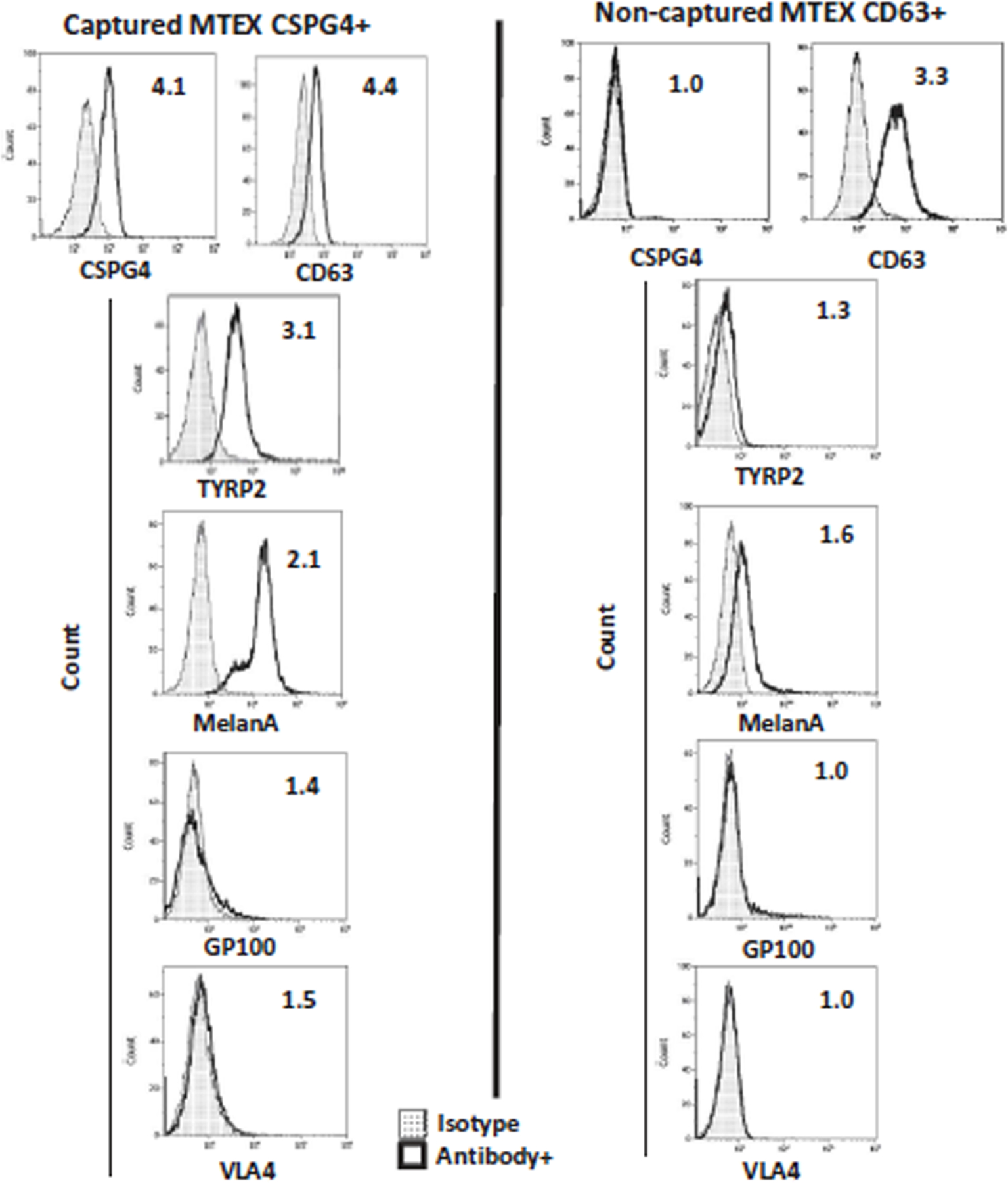
Fig. 5:

Representative on bead flow cytometry for detection of melanoma-associated antigens (MAAs) on the immunocaptured MTEX and NMTEX from plasma of a melanoma patient. NMTEX were re-captured on beads with biotinylated anti-CD63 mAb. The values within the histogram represent relative fluorescence values (RFI). RFI = MFI of detection Ab/MFI of isotype control Ab. Note that MAAs are carried exclusively by CSPG4(+) MTEX and are absent from non-captured exosomes (NMTEX). The figure is adapted from Sharma et al. [16] and reproduced with permission of the Taylor & Francis Group.
