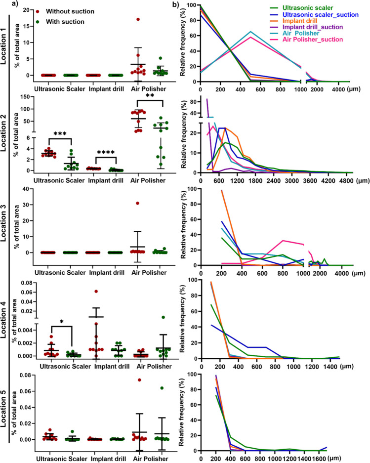
Fig. 3.
Quantification of splatter spread with/without HVS expressed as a percentage of the total area (a) and the histogram patterns of particle size (b). a Splatters quantification by calculating the % of the total area of each filter paper for every dental AGP with and without HVS for 5 locations. Each dot indicates an independent experiment. The HVS significantly reduced splatters for all three dental AGPs at location 2. At location 4, HVS significantly decreased splatter particles for ultrasonic scaler. Data are displayed as mean values ± SD. *p < 0.05; **p < 0.002; ***p < 0.0002; ****p < 0.0001 between with HVS and without HVS. b The use of HVS did not change the histogram of splatter particle size for all locations. For locations 1–3, splatter particles peaked at ~ 600 μm, while particles were smaller (peaked at 300–400 μm) at locations 4–5. This indicates large particles mainly deposit at the dentist, dental assistant, and patient’s chest

