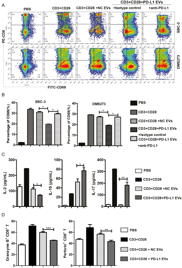
Figure 3:
EVs containing PD-L1+ downregulate CD69 expression on effector CD8+ T cells. (A) FACS contour plots showing a frequency of CD8+CD69+ cells in total CD8+ T cells in the PBMCs following various treatments at day 2. Decreasing levels of CD69 expression on CD8+ T cells after co-incubation with PD-L1+ EVs isolated from SBC-3-PD-L1 and DMS273-PD-L1 cells. The results are representative of three independent experiments. (B) Statistical analysis of the difference in expression of CD69 by flow cytometry in CD8+ T cells. ∗P < 0.05. (C) Multi-microsphere flow immunofluorescence assay assessment for secretion of IL-2, IL-17, and IL-10 in human PBMCs (stimulated with anti-CD3/CD28 antibodies) after treatment with SBC-3-NC-derived EVs or SBC-3-PD-L1 cell-derived EVs for 2 days. (D) PBMCs were analyzed by flow cytometry after staining with anti-human antibodies CD8-APC, granzyme B-PE, and perforin-FITC. Histograms represent the mean ± SD of three independent experiments. ∗∗P < 0.01. ∗∗∗P < 0.001.

