
Figure 5. Modulation of Integrin β1 signaling by Lgl1 is essential for epithelial migration.
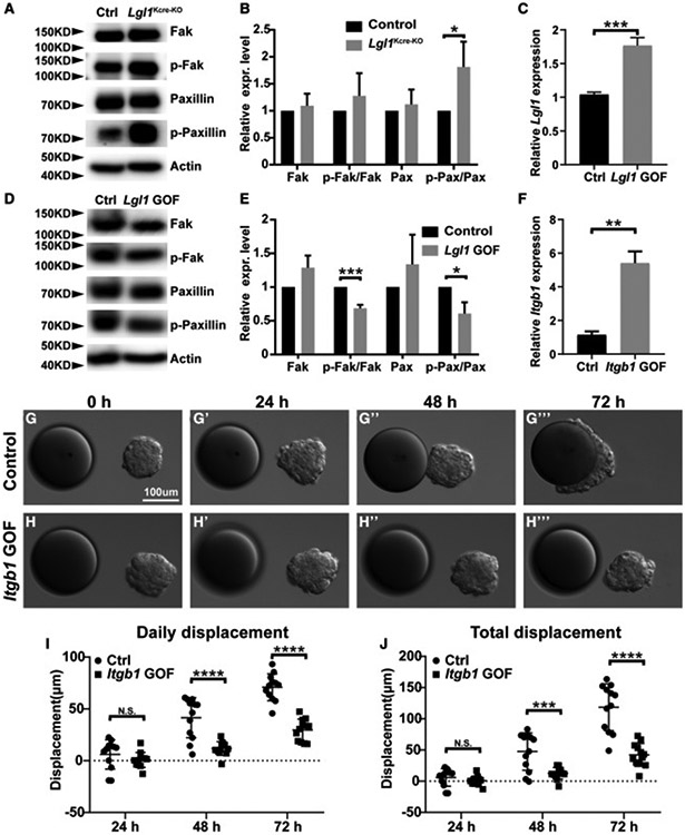
(A and B) Effect of Lgl1 loss on Integrin β1 signaling activation, as measured by the phosphorylation status of Integrin β1 downstream components Fak and Paxillin using western blot analysis. (A) Assays were performed using control and Lgl1 null MECs. (B) Relative levels of active Fak and Paxillin as measured by the ratio of phosphorylated and total forms of Fak and Paxillin.
(C–E) Effect of Lgl1 overexpression on Fak and Paxillin phosphorylation in the HC11 cell line using western blot analysis. (C) Relative levels of Lgl1 overexpression were measured by a qPCR analysis. (D) Assays were performed using control and Lgl1 GOF HC11 cells. (E) Relative levels of active Fak and Paxillin as measured by the ratio of phosphorylated and total forms of Fak and Paxillin.
(F–J) Effect of Itgb1 overexpression on collective migration using the HC11 cell line. (F) Relative levels of Itgb1 overexpression were measured by a qPCR analysis. Time course of control (G–G‴) and Itgb1 GOF (H–H‴) HC11 cell aggregate migration toward beads pre-soaked in FGF10. Quantification of the daily (I) and total (J) displacement of HC11 cells with and without Itgb1 overexpression.
Data are mean ± SD. Statistical analysis was performed using unpaired Student’sttest. *p < 0.05; **p < 0.01; ***p < 0.001; ****p< 0.0001; N.S., not significant. See also Figure S5.