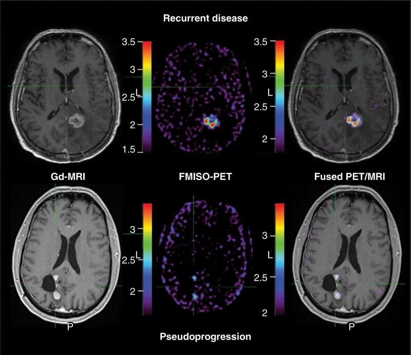
Figure 1.
FMISO PET MRI at the time of glioblastoma presumed disease progression. Gadolinium-enhanced MRI (Gd-MRI; left) at the time of presumed disease progression in patients treated with Stupp protocol with concurrent pembrolizumab demonstrates similar appearing contrast-enhancing mass in patients with both recurrent tumor growth (top) and neuroinflammatory-based therapeutic changes; pseudoprogression (PSP, below). Unlike recurrent disease, patients with PSP demonstrated minimal hypoxic volume (middle column) and hypoxic fraction (right column; ratio of hypoxic volume to gadolinium-enhancing volume). In this example, the patient with recurrent disease demonstrated a hypoxic disease burden of 178%. Conversely, the example of PSP demonstrated a hypoxic fraction of 24%. Note: FMISO PET image (middle) window minimum is the mean cerebellar background and window maximum is 2× the mean cerebellar background. Fused PET/MRI image (right) window is the hypoxic volume of 1.2× the mean cerebellar background and window maximum is 2× the hypoxic volume SUV. PET intensity color scales represent the respective visualized SUV measures. Abbreviations: FMISO, [18F]-fluoromisonidazole; MRI, magnetic resonance imaging; PET, positron emission tomography; SUV, standard uptake values.

