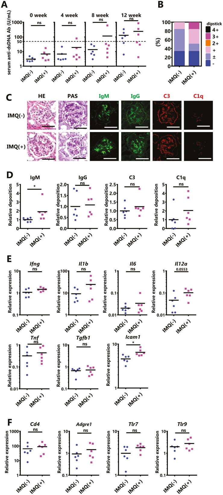
Figure 5.
Worsening of nephritis by IMQ requires a predisposition to SLE. IMQ was topically administered to 20-week-old male BWF1 mice three times weekly, similarly to females, on one ear for 12 weeks. (A) The anti-dsDNA Ab level in serum was measured by an ELISA before treatment, and after 4, 8, or 12 weeks of IMQ treatment. (N = 6 mice per group). (B) Proteinuria was measured by dipstick at sampling. The ratio of each score is shown. (N = 6 mice per group). (C) Representative images of kidney sections from untreated BWF1 mice or BWF1 mice treated with topical IMQ for 12 weeks. Sections were stained with HE, PAS, anti-IgM, anti-IgG, anti-C3, and anti-C1q. Bar = 50 μm. (D) The deposition of IgM, IgG, C3, and C1q in the kidney is shown using the mean fluorescence intensity (N = 6 mice per group). (E, F) Whole kidneys were harvested from these mice, and RNA was extracted. The expression of (E) Ifng, Il1b, Il6, Il12a, Tnf, Tgfb1, and Icam1. (F) Cd4, Adgre1 (also known as F4/80), Tlr7, and Tlr9 was determined by qPCR. The expression of the indicated genes was normalized to that of Actb. All graphs show the mean values, and each dot shows an individual mouse (N = 6 mice per group). The results were analyzed by a two-tailed Student’s t-test. Asterisks indicate statistically significant differences (∗P < 0.05, ∗∗P < 0.01, ns: not significant) and numbers indicate P values.

