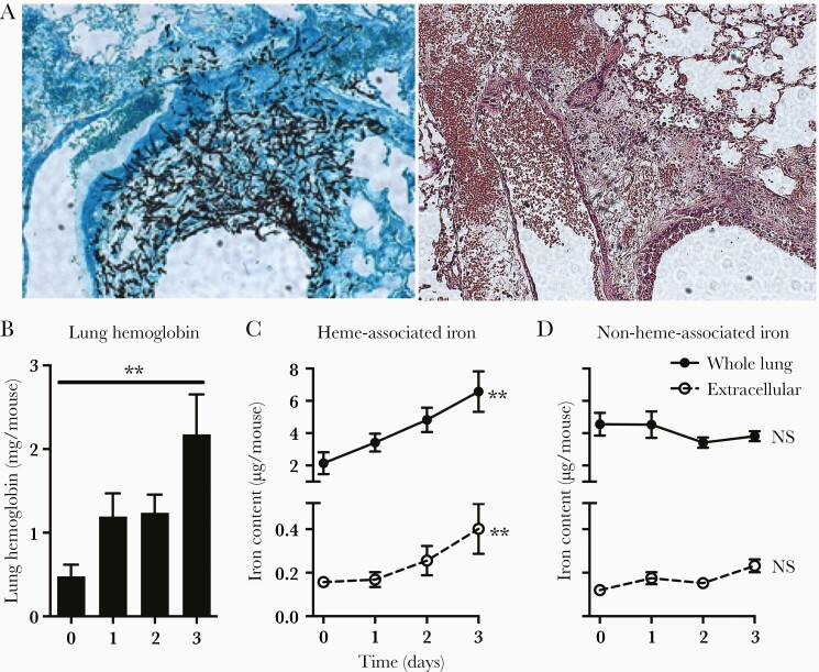
Figure 1.
Lung hemorrhage during invasive pulmonary aspergillosis. (A) Lung histology on day 2 of infection. Serial sections of the lung show invasion of a pulmonary venule by fungal hyphae from an adjacent airway and alveoli (left, Gomori's methenamine silver stain), and hemorrhage into the lung parenchyma (right, hematoxylin and eosin stain). Original magnification ×100. (B) Hemoglobin concentration in whole lung homogenates. (C and D) Heme- and nonheme-associated iron in whole lungs (intra- and extracellular) and supernatant of bronchoalveolar lavage fluid (extracellular). Data represent mean ± standard error of the mean of n = 5–10 animals per time point in each panel. **, P < .01; time 0 represents neutropenic but uninfected animals. NS, no significant difference.

