Figure 3.
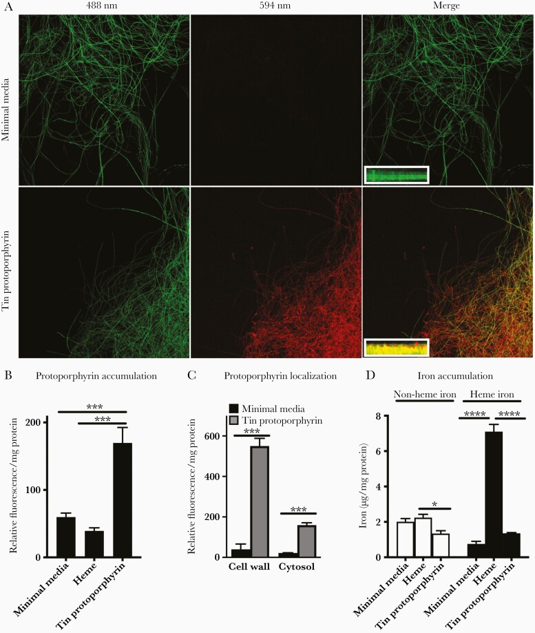
Aspergillus uptake of heme and the heme analog, tin-protoporphyrin. (A) Representative confocal images of a green fluorescent protein (GFP)-expressing Aspergillus fumigatus hyphae cultured in minimal media or minimal media supplemented with tin(IV) protoporphyrin, obtained with indicated laser emission wavelengths, and 2D Z-stack images of hyphal fragments (insets, 0.5-μm steps) showing colocalization of GFP (green) and tin-protoporphyrin fluorescence (red) in the fungal cytoplasm. Original magnifications ×200 for main panels and ×630 for insets. (B and C) Fluorescence at excitation/emission of 425/590 nm normalized to total protein of A fumigatus cultured for 2 days in indicated media. (D) Heme- and nonheme iron recovered from the cytosolic fraction normalized to cytosolic protein content of A fumigatus cultured for 2 days in indicated media. Data represent mean ± standard error of the mean of n = 3–6 replicates in each panel. *, ***, and **** denote P < .05, P < .001, and P < .0001, respectively.

