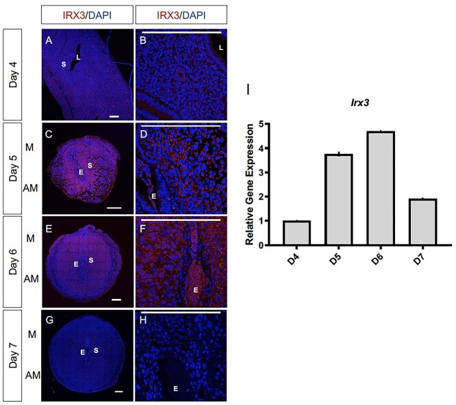
Figure 2.
Irx3 expression coincides with embryo implantation. (A–F) Immunofluorescence of IRX3 (red) co-labeled with DAPI, a nuclear marker (blue) throughout pregnancy days 4–7 (D4–D7) in wild type mice implantation sites (n = 3). Scale bars represent 250 μm. L: lumen; S: stroma; E: embryo; M: mesometrial; AM: anti-mesometrial. (I) Characterization of Irx3 transcripts in the WT pregnant mouse uterus at D4-7. Real-time qPCR was determined by setting the expression level of Irx3 mRNA on D4 of pregnancy to 1.0. Results are reported relative to 36b4 (n = 3). Data represent the mean ± SEM of three biological replicates performed in triplicate at each time point.

