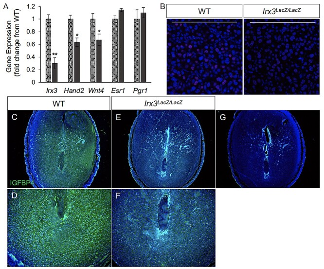
Figure 4.
Decidualization is impaired in pregnant Irx3LacZ/LacZ uteri. (A) Real-time qPCR was performed using total RNA isolated from implantation sites from pregnant uteri of WT (stippled light gray) and Irx3LacZ/LacZ (dark gray) females on day 7 of pregnancy. Data represent the mean ± SEM of three biological replicates performed in triplicate at each time point. Fold change was calculated relative to transcript levels of the WT sample. Statistics: Student’s t-test, *P < 0.05. (B) Immunofluorescence of HAND2, a stromal cell marker (red) co-localized with DAPI, a nuclear marker (blue) at gestation D7 in control (WT, n = 4) and Irx3LacZ/LacZ (n = 3) implantation sites. Scale bars represent 250 mm. (C–F) Immunofluorescence of IGFBP4 (green, DAPI in blue), a marker for decidualized stromal cells at D7 in control (C, D) versus Irx3LacZ/LacZ (E, F) embryonic implantation sites. (D, F) Magnified views of the antimesometrial zone of the implantation sites for control (D) and Irx3LacZ/LacZ (F). (G) No antibody control.

