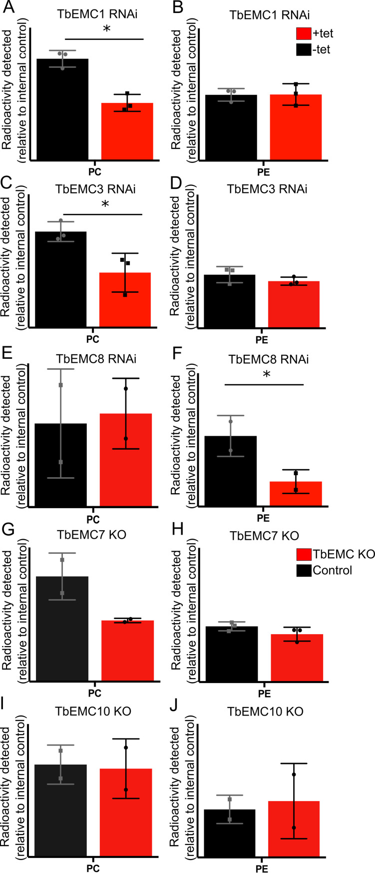
Fig 10. Analysis of [3H]-PC and [3H]-PE formation in TbEMC-depleted parasites.
Parasites cultured in the absence (black) or the presence (red) of tetracycline to maintain or ablate, respectively, TbEMC expression (A-F), or parasites lacking individual TbEMC proteins (G-J), were labeled with [3H]-choline or [3H]-ethanolamine, in combination with [3H]-inositol as internal standard, to measure de novo formation of phosphatidylcholine (PC) or phosphatidylethanolamine (PE), respectively. After extraction, phospholipids were separated by TLC and the amounts of radioactivity in individual peaks were quantified by radioisotope scanning. The data points are from independent experiments and are expressed relative to control values (means ± standard deviations). The asterisks indicate statistical significance (P<0.05).

