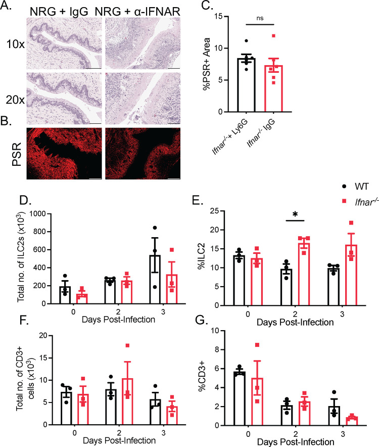
Fig 2. Vaginal immunopathology in Ifnar-/- mice is independent of ILC2s or TH2 CD4+ T cells.
(A) H&E staining of vaginal cross-sections of NRG mice + α-IFNAR or isotype control 3 dpi (n = 5). (B and C) PSR staining (B) and quantification of PSR+ to total vaginal tissue (C) of NRG mice + α-IFNAR or isotype control 3 dpi (n = 5). (D and E) Total number of CD45+Lin-ST2+CD90.2+ ILC2s (D) and proportion of ILC2s to total CD45+ cells (E) in vaginal tissue of WT and Ifnar-/- mice following HSV-2 infection (n = 3). (F and G) Total number of CD3+ T cells (F) and proportion of CD3+T cells to total CD45+ cells (G) in vaginal tissue (n = 3). 10x scale bar represents 200 μm, 20x and PSR scale bar represents 100 μm. Data in (C) to (D), (E), (F), and (G), are represented as mean ± SEM. *p < 0.05. (D-G, two-way ANOVA; C two-tailed t test) See also S2 Fig.

