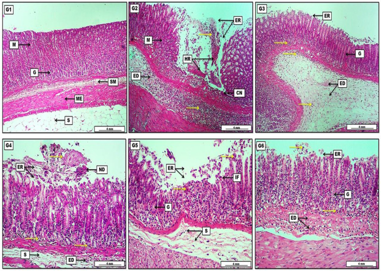
Figure 6.
Photomicrograph of gastric tissue from groups; (G1) Shows no obvious lesions, represented by intact and typically arranged gastric layers started with the mucosa (M) with normally appearing gastric glands (G), then a loose connective tissue of submucosa (SM), two layers of smooth muscle representing the muscularis externa (ME), and finally the serosa (S) which consist of adipose tissue. (G2) Reveal significant area of mucosal erosion (ER) together with the presence of hemorrhage (HR) mixed with necrotic tissue, and diffusely distributed inflammatory exudates (yellow arrows) throughout the mucosal (M) erosion and the submucosa which contains also a pinkish edematous fluid (ED). (G3) Elucidate the presence of light pinkish edema (ED) within the submucosa mixed with inflammatory exudates (yellow arrows). The inflammatory exudates can also be seen among the gastric glands (G) which show a regenerative capacity in the area of gastric mucosal erosion (ER). (G4) Show pinkish-proteinaceous inflammatory exudates (yellow arrows) mixed with mucosal necrotic debris (ND) in the area of mucosal erosion (ER). The inflammatory exudates are also distributed among the gastric glands, together with the presence of submucosal (S) inflammatory edema (ED). (G5) Display slight mucosal necrotic debris (yellow arrows) in the area of gastric erosion (ER) mixed with some inflammatory cells (IF). Regeneration of some damaged gastric glands (G) and some edema with a scant number of inflammatory cells in the submucosa (S). (G6) show clear regeneration of mucosal gastric glands (G), reduction in the area of mucosal erosion (ER) together with few inflammatory cell infiltration (yellow arrows), and pinkish inflammatory edema (ED) can be noted clearly within the submucosal layer. H&E. Scale bar: 4 mm.

