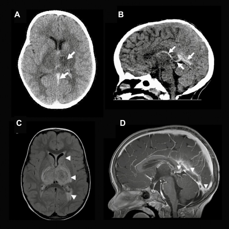
Figure 1.
Brain images of 2-year-old girl with iron deficiency anemia, demonstrating cerebral sinus venous thrombosis. CT head demonstrates a hypodense left thalamus, a high attenuating density present within the straight sinus (SS), favored vein of Galen (VoG), and internal cerebral vein consistent with CVST, seen in the axial (Panel A) and sagittal views (Panel B). (Arrows). (Panel C) The MRI FLAIR-weighted axial image depicts increased signal in the bilateral thalamus, globus pallidus, genu and posterior limbs of the internal capsule, putamen, and the trigones suggestive of extensive edema. Also, a mild ventriculomegaly is evident. (Panel D) The sagittal enhanced T1-weighted image shows the intraluminal thrombus filling the torcula, SS, and VoG (Arrow heads).

